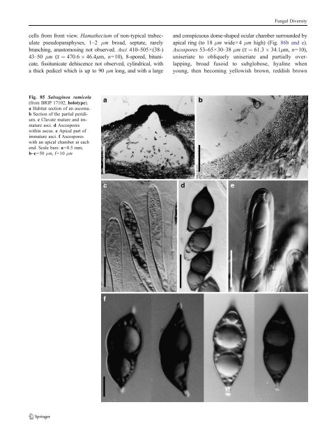
Fungal Diversity cells from front view. Hamathecium of non-typical trabeculate pseudoparaphyses, 1–2 μm broad, septate, rarely branching, anastomosing not observed. Asci 410–505×(38-) 43–50 μm (x ¼ 470:6 46:4mm, n=10), 8-spored, bitunicate, fissitunicate dehiscence not observed, cylindrical, with a thick pedicel which is up to 90 μm long, and with a large and conspicuous dome-shaped ocular chamber surrounded by apical ring (to 18 μm wide×4 μm high) (Fig. 86b and e). Ascospores 53–65×30–38 μm (x ¼ 61:3 34:1mm, n=10), uniseriate to obliquely uniseriate and partially overlapping, broad fusoid to subglobose, hyaline when young, then becoming yellowish brown, reddish brown Fig. 85 Salsuginea ramicola (from BRIP 17102, holotype). a Habitat section of an ascoma. b Section of the partial peridium. c Clavate mature and immature asci. d Ascospores within ascus. e Apical part of immature asci. f Ascospores with an apical chamber at each end. Scale bars: a=0.5 mm, b–e=50 μm, f=10μm
Fungal Diversity and nearly black and opaque when mature, non-septate, smooth-walled, with a full length germ slit, surrounded by a broad gelatinous sheath (Fig. 86c and d). Anamorph: none reported. Material examined: CANADA, Alberta, North of Beaver Mines, on sheep dung, 28 Jul. 1962, E.R. Luck- Allen, (TRTC 41607, paratype); USA, Montana: Gallatin County, 60 min S of Bozeman, on sheep dung, 2 Sept. 1957, Cain (TRTC 42032, paratype); Stillwater County Columbus, on cow dung, 3 Sept. 1957, Cain (TRTC 42031, paratype); South Dakota, Meade Co.: South of Wall, on cow dung, 3 Sept. 1962, Cain (TRTC 40697, holotype). Notes Morphology Semidelitschia was formally establishedbyCainandLuck- Allen (1969) and was assigned to Sporormiaceae. Although it is similar to Delitschia, it differs as the ascospores are 1- celled, as opposed to 2-celled. Subsequently, Semidelitschia was transferred to Delitschiaceae together with Delitschia (Barr 2000). Currently, three species are listed under this genus, i.e. S. agasmatica Cain & Luck-Allen, S. nanostellata A.E. Bell & Mahoney and S. tetraspora J.H. Mirza & S.M. Khan (Index Fungorum) although the number of species in the genus are given as only two in Kirk et al. (2008). Phylogenetic study None. Concluding remarks This is a clearly defined genus that differs from Delitschia in having 1-celled ascospores. Cultures of S. agasmatica are needed for sequencing and for establishing the placement and uniqueness of the genus. Setomelanomma M. Morelet, Bull. Soc. Sci. nat. Arch. Toulon et du Var 227:15 (1980). (Phaeosphaeriaceae) Generic description Habitat terrestrial, hemibiotrophic or biotrophic. Ascomata small, solitary, scattered, immersed, erumpent to superficial, globose to subglobose, black; with or without a small papilla, apex covered with setae and a periphysate ostiole. Peridium thin, 1-layered, composed of several layers of cells of textura angularis. Hamathecium of dense, 1–2 μm broad pseudoparaphyses, septate, anastomosing. Asci 8- spored, bitunicate, broadly cylindrical. Ascospores fusoid to broadly clavate, pale brown to brown, 3-septate. Anamorphs reported for genus: none. Literature: Leonard and Suggs 1974; Morelet 1980; Rossman et al. 2002; Schochetal.2009; Zhang et al. 2009a. Type species Setomelanomma holmii M. Morelet, Bulletin de la Société des Sciences naturelles et d’Archéologie de Toulon et du Var 36 (no. 227): 15 (1980). (Fig. 87) (Some information in the following description is from Rossman et al. (2002)) Ascomata 80–250 μm diam., solitary, scattered, immersed, erumpent to superficial, globose to subglobose, black, with setae; with or without a small papilla, apex covered with setae and a periphysate ostiole. Peridium 15– 25 μm thick, 1-layered, composed of several layers of cells of textura angularis, cell wall thinner and more lightly pigmented towards centrum, cell wall thicker near the apex. Hamathecium of dense, 1–2 μm broad pseudoparaphyses, thicker near the base, septate, anastomosing (Fig. 87a and d). Asci 70–100×11–14 μm, 8-spored, bitunicate, broadly cylindrical with a short, thick, furcate pedicel, with a small ocular chamber (Fig. 87a, b and c). Ascospores 16–21×5– 6.5 μm, obliquely uniseriate and partially overlapping to biseriate, fusoid to broadly clavate with broadly to narrowly rounded ends, pale brown to brown, 3-septate, slightly constricted at the median septum, smooth (Fig. 87e). Anamorph: none reported. Material examined: FRANCE, Leuglay, on dying twigs of Picea pungens. 8 May 1987, leg. M. Morelet (UPS F- 117969 (slide), isotype). Notes Morphology Setomelanomma was formally established by Morelet (1980) as a monotypic genus represented by S. holmii, which was collected in France. The description, however, is not detailed and lacks illustrations. Rossman et al. (2002) collected this species in North America and detailed studies were conducted including both morphology and phylogeny. The bitunicate, broadly cylindrical asci, cellular pseudoparaphyses as well as the pale brown, septate ascospores with a median primary septum point Setomelanomma to Phaeosphaeriaceae as defined by Barr (1992a) and Eriksson et al. (2002) (Rossman et al. 2002). However, its setose ascomata, brown and 3-septate ascospores together with its residence in conifers distinguish it from all other genera under Phaeosphaeriaceae (Rossman et al. 2002). Setomelanomma is mostly comparable with Kalmusia and Phaeosphaeria. Setomelanomma can be distinguished from Kalmusia by its erumpent to superficial ascomata with almost no papilla, and Phaeosphaeria differs from Setomelanomma by its host spectrum and reported anamorphic stages (Rossman et al. 2002). Currently, five species are included in Setomelanomma, namely S. holmii, S. monoceras, S. prolata K.J. Leonard & Suggs, S. rostrata (K.J. Leonard) K.
- Page 1 and 2:
Fungal Diversity DOI 10.1007/s13225
- Page 3 and 4:
Fungal Diversity Table 1 Major circ
- Page 5 and 6:
Fungal Diversity
- Page 7 and 8:
Fungal Diversity biocontrol agent o
- Page 9 and 10:
Fungal Diversity substrates and man
- Page 11 and 12:
Fungal Diversity 2. To investigate
- Page 13 and 14:
Fungal Diversity Table 3 (continued
- Page 15 and 16:
Fungal Diversity Table 3 (continued
- Page 17 and 18:
Fungal Diversity Table 3 (continued
- Page 19 and 20:
Fungal Diversity
- Page 21 and 22:
Fungal Diversity Fig. 2 Aigialus gr
- Page 23 and 24:
Fungal Diversity Fig. 3 Amniculicol
- Page 25 and 26:
Fungal Diversity Literature: Berkel
- Page 27 and 28:
Fungal Diversity Ascorhombispora L.
- Page 29 and 30:
Fungal Diversity
- Page 31 and 32:
Fungal Diversity Fig. 8 Astrosphaer
- Page 33 and 34:
Fungal Diversity Fig. 9 Asymmetrico
- Page 35 and 36:
Fungal Diversity Notes Morphology B
- Page 37 and 38:
Fungal Diversity Generic descriptio
- Page 39 and 40:
Fungal Diversity Anamorph: none rep
- Page 41 and 42:
Fungal Diversity Fig. 14 Bimuria no
- Page 43 and 44:
Fungal Diversity Fig. 15 Bricookea
- Page 45 and 46:
Fungal Diversity Fig. 16 Byssolophi
- Page 47 and 48:
Fungal Diversity Notes Morphology B
- Page 49 and 50:
Fungal Diversity the reaction of pe
- Page 51 and 52:
Fungal Diversity
- Page 53 and 54:
Fungal Diversity Fig. 21 Chaetomast
- Page 55 and 56:
Fungal Diversity
- Page 57 and 58:
Fungal Diversity Fig. 23 Cilioplea
- Page 59 and 60:
Fungal Diversity with one or two ve
- Page 61 and 62:
Fungal Diversity Moreau 1953; Munk
- Page 63 and 64:
Fungal Diversity Material examined:
- Page 65 and 66:
Fungal Diversity Fig. 28 Dothidotth
- Page 67 and 68:
Fungal Diversity Fig. 29 Dubitatio
- Page 69 and 70:
Fungal Diversity assigned Entodesmi
- Page 71 and 72:
Fungal Diversity fusoid to somewhat
- Page 73 and 74:
Fungal Diversity Fig. 33 Hadrospora
- Page 75 and 76:
Fungal Diversity Fig. 34 Halotthia
- Page 77 and 78:
Fungal Diversity Notes Morphology H
- Page 79 and 80:
Fungal Diversity some effused Hypox
- Page 81 and 82:
Fungal Diversity Fig. 38 Isthmospor
- Page 83 and 84:
Fungal Diversity Fig. 39 Kalmusia e
- Page 85 and 86:
Fungal Diversity ascospores were br
- Page 87 and 88:
Fungal Diversity furcate pedicel an
- Page 89 and 90:
Fungal Diversity Anamorph: none rep
- Page 91 and 92:
Fungal Diversity
- Page 93 and 94:
Fungal Diversity Material examined:
- Page 95 and 96:
Fungal Diversity Fig. 46 Lewia scro
- Page 97 and 98:
Fungal Diversity Fig. 47 Lichenopyr
- Page 99 and 100:
Fungal Diversity Loculohypoxylon M.
- Page 101 and 102:
Fungal Diversity cells small heavil
- Page 103 and 104:
Fungal Diversity upper place, septa
- Page 105 and 106:
Fungal Diversity
- Page 107 and 108:
Fungal Diversity (CBS 627.86) was i
- Page 109 and 110: Fungal Diversity Fig. 54 Mamillisph
- Page 111 and 112: Fungal Diversity Fig. 55 Massarina
- Page 113 and 114: Fungal Diversity phaeria as a synon
- Page 115 and 116: Fungal Diversity 5-8 μm diam., ind
- Page 117 and 118: Fungal Diversity cell wall
- Page 119 and 120: Fungal Diversity Fig. 60 Mixtura sa
- Page 121 and 122: Fungal Diversity Fig. 61 Montagnula
- Page 123 and 124: Fungal Diversity spored, bitunicate
- Page 125 and 126: Fungal Diversity Fig. 64 Murispora
- Page 127 and 128: Fungal Diversity Type species Neoph
- Page 129 and 130: Fungal Diversity brown, 8-septate,
- Page 131 and 132: Fungal Diversity Fig. 68 Ohleria mo
- Page 133 and 134: Fungal Diversity Fig. 69 Ohleriella
- Page 135 and 136: Fungal Diversity Fig. 70 Ophiobolus
- Page 137 and 138: Fungal Diversity Type species Ostro
- Page 139 and 140: Fungal Diversity
- Page 141 and 142: Fungal Diversity (Shoemaker and Bab
- Page 143 and 144: Fungal Diversity ium thin, composed
- Page 145 and 146: Fungal Diversity Fig. 76 Platysporo
- Page 147 and 148: Fungal Diversity Fig. 77 1 Pleomass
- Page 149 and 150: Fungal Diversity Fig. 78 Pleophragm
- Page 151 and 152: Fungal Diversity papillate, ostiola
- Page 153 and 154: Fungal Diversity Williams 1963; Mal
- Page 155 and 156: Fungal Diversity Generic descriptio
- Page 157 and 158: Fungal Diversity composed of one ce
- Page 159: Fungal Diversity Fig. 84 Saccharico
- Page 163 and 164: Fungal Diversity dense, long trabec
- Page 165 and 166: Fungal Diversity
- Page 167 and 168: Fungal Diversity
- Page 169 and 170: Fungal Diversity Anamorphs reported
- Page 171 and 172: Fungal Diversity
- Page 173 and 174: Fungal Diversity
- Page 175 and 176: Fungal Diversity Fig. 94 Westerdyke
- Page 177 and 178: Fungal Diversity Fig. 95 Wettsteini
- Page 179 and 180: Fungal Diversity Fig. 96 Wilmia bra
- Page 181 and 182: Fungal Diversity Current name: Astr
- Page 183 and 184: Fungal Diversity spores are actuall
- Page 185 and 186: Fungal Diversity Fig. 100 Sporormie
- Page 187 and 188: Fungal Diversity
- Page 189 and 190: Fungal Diversity Fig. 102 Kriegerie
- Page 191 and 192: Fungal Diversity Phylogenetic study
- Page 193 and 194: Fungal Diversity Fig. 104 Zeuctomor
- Page 195 and 196: Fungal Diversity Fig. 105 Muroia ni
- Page 197 and 198: Fungal Diversity pseudoparenchymato
- Page 199 and 200: Fungal Diversity Eremodothis Arx, K
- Page 201 and 202: Fungal Diversity Type species: Macr
- Page 203 and 204: Fungal Diversity ascospores of Plat
- Page 205 and 206: Fungal Diversity monoceras Alcorn n
- Page 207 and 208: Fungal Diversity tomataceae, Melano
- Page 209 and 210: Fungal Diversity Table 4 (continued
- Page 211 and 212:
Fungal Diversity 1987b). Based on a
- Page 213 and 214:
Fungal Diversity only do so under v
- Page 215 and 216:
Fungal Diversity Dennis RWG (1968)
- Page 217 and 218:
Fungal Diversity Kirk PM, Cannon PF
- Page 219 and 220:
Fungal Diversity Saccardo PA (1880)
- Page 221:
Fungal Diversity Winter G (1887) As