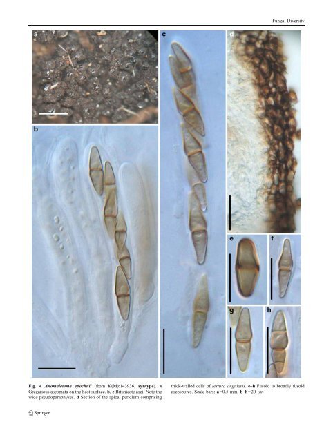
Fungal Diversity Fig. 4 Anomalemma epochnii (from K(M):143936, syntype). a Gregarious ascomata on the host surface. b, c Bitunicate asci. Note the wide pseudoparaphyses. d Section of the apical peridium comprising thick-walled cells of textura angularis. e–h Fusoid to broadly fusoid ascospores. Scale bars: a=0.5 mm, b–h=20 μm
Fungal Diversity Literature: Berkeley and Broome 1866; Keissler 1922;Massee 1887; Saccardo 1878a; Sivanesan 1983. Type species Anomalemma epochnii (Berk. & Broome) Sivan., Trans. Br. Mycol. Soc. 81: 328 (1983). (Fig. 4) ≡ Sphaeria epochnii Berk. & Broome, Ann. Mag. nat. Hist., Ser. 3 18: 128 (1866). Ascomata 340–500 μm high×170–286 μm diam., gregarious on the intertwined hyphae, superficial, papillate, wall black, coriaceous, roughened (Fig. 4a). Peridium composed of two types of cells, outer layer 17–22 μm wide, composed of heavily pigmented thick-walled cells of textura angularis, cells up to 8×13 μm diam., cell wall 1–1.5 μm thick, inner layer 30–34 μm thick, composed of hyaline thin-walled cells (Fig. 4d). Hamathecium of dense, long cellular pseudoparaphyses, 2–4 μm broad, septate. Asci 75–108×9.5–12.5 μm (x ¼ 92:8 11:1mm, n=10), 8-spored, bitunicate, fissitunicate, dehiscence not observed, cylindro-clavate to clavate, with a furcate pedicel up to 6–25 μm long, with a small ocular chamber best seen in immature asci (ca. 2μm wide×1 μm high) (Fig. 4b and c). Ascospores 20–25(−30)×5–7.5 μm (x ¼ 23:1 6:3mm, n=10), obliquely uniseriate and partially overlapping to biseriate, fusoid to narrowly fusoid with narrowly rounded ends, brown, 1-septate, rarely 2- to 3-septate, deeply constricted at the median septum, smooth (Fig. 4e, f, g and h). Anamorph: Exosporiella fungorum (Fr.) P. Karst. (Sivanesan 1983). = Epochnium fungorum Fr., Syst. mycol. 3: 449 (1832). Mycelium composed of branched, septate, pale brown hyphae. Stroma none. Conidiophores macronematous or semi-macronematous, mononematous, hyaline, smooth, branched towards the apex. Conidiogenous cells monoblastic, cylindrical or doliform. Conidia cylindrical or ellipsoidal, dry, 3-4-septate, smooth, hyaline or pale brown. Material examined: UK, England, Warleigh near Bath, on fungus on bark (Epochnium sp.), Mar. 1866, leg. Warbright? (K(M):143936, syntype, ex herb. C.E. Broome). Notes Morphology Sphaeria epochnii was first described and illustrated by Berkeley and Broome (1866) from Britain and the anamorphic stage is the hyphomycetous Epochniella fungorum. Sphaeria epochnii has subsequently been transferred to Melanomma (as M. epochnii (Berk. & Broome) Sacc.; Saccardo 1878a), Byssosphaeria (as B. epochnii (Berk. & Broome) Cooke; Massee 1887) andChaetosphaeria (as C. epochnii (Berk. & Broome) Keissl.; Keissler 1922). The deposition of Sphaeria epochnii in Chaetosphaeria is obviously unacceptable, as Chaetosphaeria has unitunicate asci. Melanomma has been reported having Aposphaeria or Pseudospiropes anamorphs, which differs from Exosporiella (Sivanesan 1983). In addition, the presence of well developed prosenchymatous stroma in Sphaeria epochnii can also readily distinguish it from Melanomma (Sivanesan 1983). The gregarious ascomata and formation of prosenchymatous stroma of Anomalemma resembles those of Cucurbitaria, but the pleosporaceous dictyosporous ascospores of Cucurbitaria readily distinguish it from Anomalemma epochnii. In addition, the pseudoparenchymatous peridium, fungicolous habitat and brown 1-septate ascospores, which later becoming 3-septate differ from any other pleosporalean genus. Thus a new genus, Anomalemma, was introduced to accommodate it (Sivanesan 1983). Anomalemma is presently monotypic. Phylogenetic study None. Concluding remarks Anomalemma epochnii certainly resembles Byssosphaeria in its ascomata clustering together in groups on closely intertwined hyphae and brown ascospores, and may well be included in this genus. Its fungicolous habitat, however, distinguishes it from Byssosphaeria. Appendispora K.D. Hyde, Sydowia 46: 29 (1994a). (?Didymellaceae) Generic description Habitat terrestrial, saprobic. Ascomata small, clustered, immersed, subglobose or irregularly pyriform. Peridium thin. Hamathecium of dense, long trabeculate pseudoparaphyses. Asci 8-spored, bitunicate, fissitunicate, cylindrical, apical rounded with ocular chamber and faint ring, with short pedicels. Ascospores uniseriate to partially overlapping, fusoid, brown, 1-septate, slightly constricted at the septum. Anamorphs reported for genus: none. Literature: Hyde 1994a. Type species Appendispora frondicola K.D. Hyde, Sydowia 46: 30 (1994a). (Fig. 5) Ascomata 120–280 μm high×180–280 μm diam., clustered, immersed with minute ostioles visible through cracks or blackened dots on the host surface, subglobose or irregularly pyriform (Fig. 5a and b). Peridium 40 μm thick, comprising two types of cells; outer cells, small heavily pigmented thick-walled cells of textura angularis, innercells compressed, hyaline. Hamathecium of dense, very long trabeculate pseudoparaphyses, ca. 1μm broad, embedded in mucilage, hyaline, anastomosing (Fig. 5e). Asci 130–144×11–
- Page 1 and 2: Fungal Diversity DOI 10.1007/s13225
- Page 3 and 4: Fungal Diversity Table 1 Major circ
- Page 5 and 6: Fungal Diversity
- Page 7 and 8: Fungal Diversity biocontrol agent o
- Page 9 and 10: Fungal Diversity substrates and man
- Page 11 and 12: Fungal Diversity 2. To investigate
- Page 13 and 14: Fungal Diversity Table 3 (continued
- Page 15 and 16: Fungal Diversity Table 3 (continued
- Page 17 and 18: Fungal Diversity Table 3 (continued
- Page 19 and 20: Fungal Diversity
- Page 21 and 22: Fungal Diversity Fig. 2 Aigialus gr
- Page 23: Fungal Diversity Fig. 3 Amniculicol
- Page 27 and 28: Fungal Diversity Ascorhombispora L.
- Page 29 and 30: Fungal Diversity
- Page 31 and 32: Fungal Diversity Fig. 8 Astrosphaer
- Page 33 and 34: Fungal Diversity Fig. 9 Asymmetrico
- Page 35 and 36: Fungal Diversity Notes Morphology B
- Page 37 and 38: Fungal Diversity Generic descriptio
- Page 39 and 40: Fungal Diversity Anamorph: none rep
- Page 41 and 42: Fungal Diversity Fig. 14 Bimuria no
- Page 43 and 44: Fungal Diversity Fig. 15 Bricookea
- Page 45 and 46: Fungal Diversity Fig. 16 Byssolophi
- Page 47 and 48: Fungal Diversity Notes Morphology B
- Page 49 and 50: Fungal Diversity the reaction of pe
- Page 51 and 52: Fungal Diversity
- Page 53 and 54: Fungal Diversity Fig. 21 Chaetomast
- Page 55 and 56: Fungal Diversity
- Page 57 and 58: Fungal Diversity Fig. 23 Cilioplea
- Page 59 and 60: Fungal Diversity with one or two ve
- Page 61 and 62: Fungal Diversity Moreau 1953; Munk
- Page 63 and 64: Fungal Diversity Material examined:
- Page 65 and 66: Fungal Diversity Fig. 28 Dothidotth
- Page 67 and 68: Fungal Diversity Fig. 29 Dubitatio
- Page 69 and 70: Fungal Diversity assigned Entodesmi
- Page 71 and 72: Fungal Diversity fusoid to somewhat
- Page 73 and 74: Fungal Diversity Fig. 33 Hadrospora
- Page 75 and 76:
Fungal Diversity Fig. 34 Halotthia
- Page 77 and 78:
Fungal Diversity Notes Morphology H
- Page 79 and 80:
Fungal Diversity some effused Hypox
- Page 81 and 82:
Fungal Diversity Fig. 38 Isthmospor
- Page 83 and 84:
Fungal Diversity Fig. 39 Kalmusia e
- Page 85 and 86:
Fungal Diversity ascospores were br
- Page 87 and 88:
Fungal Diversity furcate pedicel an
- Page 89 and 90:
Fungal Diversity Anamorph: none rep
- Page 91 and 92:
Fungal Diversity
- Page 93 and 94:
Fungal Diversity Material examined:
- Page 95 and 96:
Fungal Diversity Fig. 46 Lewia scro
- Page 97 and 98:
Fungal Diversity Fig. 47 Lichenopyr
- Page 99 and 100:
Fungal Diversity Loculohypoxylon M.
- Page 101 and 102:
Fungal Diversity cells small heavil
- Page 103 and 104:
Fungal Diversity upper place, septa
- Page 105 and 106:
Fungal Diversity
- Page 107 and 108:
Fungal Diversity (CBS 627.86) was i
- Page 109 and 110:
Fungal Diversity Fig. 54 Mamillisph
- Page 111 and 112:
Fungal Diversity Fig. 55 Massarina
- Page 113 and 114:
Fungal Diversity phaeria as a synon
- Page 115 and 116:
Fungal Diversity 5-8 μm diam., ind
- Page 117 and 118:
Fungal Diversity cell wall
- Page 119 and 120:
Fungal Diversity Fig. 60 Mixtura sa
- Page 121 and 122:
Fungal Diversity Fig. 61 Montagnula
- Page 123 and 124:
Fungal Diversity spored, bitunicate
- Page 125 and 126:
Fungal Diversity Fig. 64 Murispora
- Page 127 and 128:
Fungal Diversity Type species Neoph
- Page 129 and 130:
Fungal Diversity brown, 8-septate,
- Page 131 and 132:
Fungal Diversity Fig. 68 Ohleria mo
- Page 133 and 134:
Fungal Diversity Fig. 69 Ohleriella
- Page 135 and 136:
Fungal Diversity Fig. 70 Ophiobolus
- Page 137 and 138:
Fungal Diversity Type species Ostro
- Page 139 and 140:
Fungal Diversity
- Page 141 and 142:
Fungal Diversity (Shoemaker and Bab
- Page 143 and 144:
Fungal Diversity ium thin, composed
- Page 145 and 146:
Fungal Diversity Fig. 76 Platysporo
- Page 147 and 148:
Fungal Diversity Fig. 77 1 Pleomass
- Page 149 and 150:
Fungal Diversity Fig. 78 Pleophragm
- Page 151 and 152:
Fungal Diversity papillate, ostiola
- Page 153 and 154:
Fungal Diversity Williams 1963; Mal
- Page 155 and 156:
Fungal Diversity Generic descriptio
- Page 157 and 158:
Fungal Diversity composed of one ce
- Page 159 and 160:
Fungal Diversity Fig. 84 Saccharico
- Page 161 and 162:
Fungal Diversity and nearly black a
- Page 163 and 164:
Fungal Diversity dense, long trabec
- Page 165 and 166:
Fungal Diversity
- Page 167 and 168:
Fungal Diversity
- Page 169 and 170:
Fungal Diversity Anamorphs reported
- Page 171 and 172:
Fungal Diversity
- Page 173 and 174:
Fungal Diversity
- Page 175 and 176:
Fungal Diversity Fig. 94 Westerdyke
- Page 177 and 178:
Fungal Diversity Fig. 95 Wettsteini
- Page 179 and 180:
Fungal Diversity Fig. 96 Wilmia bra
- Page 181 and 182:
Fungal Diversity Current name: Astr
- Page 183 and 184:
Fungal Diversity spores are actuall
- Page 185 and 186:
Fungal Diversity Fig. 100 Sporormie
- Page 187 and 188:
Fungal Diversity
- Page 189 and 190:
Fungal Diversity Fig. 102 Kriegerie
- Page 191 and 192:
Fungal Diversity Phylogenetic study
- Page 193 and 194:
Fungal Diversity Fig. 104 Zeuctomor
- Page 195 and 196:
Fungal Diversity Fig. 105 Muroia ni
- Page 197 and 198:
Fungal Diversity pseudoparenchymato
- Page 199 and 200:
Fungal Diversity Eremodothis Arx, K
- Page 201 and 202:
Fungal Diversity Type species: Macr
- Page 203 and 204:
Fungal Diversity ascospores of Plat
- Page 205 and 206:
Fungal Diversity monoceras Alcorn n
- Page 207 and 208:
Fungal Diversity tomataceae, Melano
- Page 209 and 210:
Fungal Diversity Table 4 (continued
- Page 211 and 212:
Fungal Diversity 1987b). Based on a
- Page 213 and 214:
Fungal Diversity only do so under v
- Page 215 and 216:
Fungal Diversity Dennis RWG (1968)
- Page 217 and 218:
Fungal Diversity Kirk PM, Cannon PF
- Page 219 and 220:
Fungal Diversity Saccardo PA (1880)
- Page 221:
Fungal Diversity Winter G (1887) As