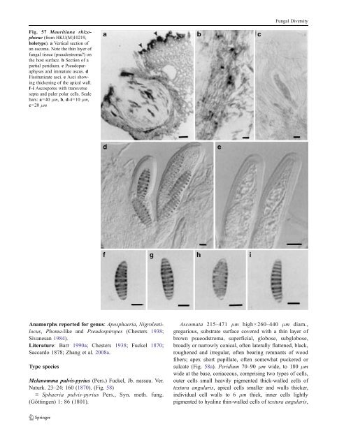
Fungal Diversity Fig. 57 Mauritiana rhizophorae (from HKU(M)10219, holotype). a Vertical section of an ascoma. Note the thin layer of fungal tissue (pseudostroma?) on the host surface. b Section of a partial peridium. c Pseudoparaphyses and immature ascus. d Fissitunicate asci. e Asci showing thickening of the apical wall. f–i Ascospores with transverse septa and paler polar cells. Scale bars: a=40 μm, b, d–i=10 μm, c=20 μm Anamorphs reported for genus: Aposphaeria, Nigrolentilocus, Phoma-like and Pseudospiropes (Chesters 1938; Sivanesan 1984). Literature: Barr 1990a; Chesters 1938; Fuckel 1870; Saccardo 1878; Zhang et al. 2008a. Type species Melanomma pulvis-pyrius (Pers.) Fuckel, Jb. nassau. Ver. Naturk. 23–24: 160 (1870). (Fig. 58) ≡ Sphaeria pulvis-pyrius Pers., Syn. meth. fung. (Göttingen) 1: 86 (1801). Ascomata 215–471 μm high×260–440 μm diam., gregarious, substrate surface covered with a thin layer of brown psueodstroma, superficial, globose, subglobose, broadly or narrowly conical, often laterally flattened, black, roughened and irregular, often bearing remnants of wood fibers; apex short papillate, often somewhat puckered or sulcate (Fig. 58a). Peridium 70–90 μm wide, to 180 μm wide at the base, coriaceous, comprising two types of cells, outer cells small heavily pigmented thick-walled cells of textura angularis, apical cells smaller and walls thicker, individual cell walls to 6 μm thick, inner cells lightly pigmented to hyaline thin-walled cells of textura angularis,
Fungal Diversity 5–8 μm diam., individual cell wall to 1.5–2 μm thick, in places with columns of textura prismatica, and larger, paler cells of textura prismatica towards the interior and at the base (Fig. 58b). Hamathecium of dense, filamentous, 1–2 (−2.5) μm broad, branching, rarely anastomosing, septate pseudoparaphyses. Asci 98–123×6.5–7.5(−9) μm (x ¼ 109 7:5mm, n=10), 8-spored, bitunicate, fissitunicate, cylindrical to fusoid, with a short, furcate pedicel, to 25 μm long, with an ocular chamber (Fig. 58c, d, e, f and g). Ascospores 14–17.5(−19)×4.5–6.5 μm (x ¼ 15:8 5:2mm, n=10), obliquely uniseriate and partially overlapping, broadly fusoid to fusoid with broadly rounded ends, straight or slightly curved, smooth, olive-brown, 4-celled, slightly constricted at the septa, the second cell from the top slightly wider than the others, no sheath (Fig. 58h,i,j,kandl). Anamorph: Aposphaeria agminalis Sacc. or Phoma agminalis Sacc. (Sivanesan 1984). Colonies (of epitype) reaching 4 cm diam. after 20 days growth on PDA at 25°C, depressed to raised, cottony to woolly, with rhizoidal margin, grey, reverse darkened. Phoma-like anamorph has been reported by Chesters (1938) and Sivanesan (1984), but no anamorphic stage was observed in the cultures of IFRDCC 2044, <strong>CBS</strong> 109.77 and <strong>CBS</strong> 371.75 after culturing 3 months on PDA. Material examined:ondecayingwood(UPS,Scler.suec. n. 120, holotype,asSphaeria pulvis-pyrius Pers.); FRANCE, Ariège, Rimont, Saurine, on bark of Salix caprea, 10Apr. 2008, Jacques Fournier (IFRD 2001, epitype). Notes Morphology Melanomma, the familial type of Melanommataceae, was formally established by Fuckel (1870, p 159) based on its small, carbonaceous ascomata, having: “sporen meist 2–3 mal septirt, selten ohne Scheidewand, braun oder wasscrhell.” (Chesters 1938; Fuckel 1870). Saccardo (1878, p. 344) emended this genus as “Spores ovate or oblong, multi-septate, coloured.” Subsequently, Saccardo (1883, p. 98) extended the description of Melanomma as “Perithecia gregarious, seldom scattered, somewhat superficial, sphaerical, papillate or blunt, carbonaceous, smooth or somewhat hairy. Asci elongate, for the most part accompanied by paraphyses, 8-spored. Spores oblong or somewhat spindle-shaped, two to many septate, olive or dark brown. Species of Sphaeria belong here for the most part.” Melanomma pulvis-pyrius was erected as the lectotype species (Barr 1990a; Chesters1938). Barr (1990a) gavea detailed circumscription for Melanomma, under which Melanomma contains about 20 species (Kirk et al. 2001). Melanomma pulvis-pyrius is characterized by its gregarious, superficial ascomata with short papillate, cylindrical asci with a short pedicel and fusoid, olive-brown, 3-septate ascospores (Chesters 1938; Zhang et al. 2008a). One of the diagnostic characters of Melanommataceae is the trabeculate pseudoparaphyses, although no typical trabeculate pseudoparaphyses could be found in the neotype (Scler. suec. n. 120, UPS) and epitype (IFRD 2001) of M. pulvis-pyrius (Zhang et al. 2008a). Phylogenetic study Phylogenetic analysis based on five genes (LSU, SSU, RPB1, RPB2 andEF1) indicates that Melanomma pulvispyrius forms a robust clade with Byssosphaeria, Herpotrichia and Pleomassaria siparia (Pleomassariaceae) and likely represents a separate family (or families comprising Melanommataceae) (Zhang et al. 2008a; Mugambi and Huhndorf 2009b). A more recent phylogenetic analysis included a group of coelomycete species with stellate conidia, isolated from Fagales trees clustered in Melanommataceae (Tanaka et al. 2010; Plate 1). Concluding remarks The Melanomma concept based on ascospore morphology appears polyphyletic. Metameris Theiss. & Syd., Annls mycol. 13: 342 (1915). (Phaeosphaeriaceae) Generic description Habitat terrestrial, saprobic or parasitic. Ascostromata erumpent through the host surface in linear rows parallel to the host fibers. Ascomata small, globose to subglobose, black, coriaceous. Peridium composed of large lightly pigmented cells of textura angularis. Hamathecium of rare, broad pseudoparaphyses, septate, constricted at the septa. Asci bitunicate, fissitunicate, broadly cylindrical to slightly obclavate, with a short, thick, knob-like pedicel. Ascospores hyaline, 1- (rarely 2-) septate. Anamorphs reported for genus: none. Literature: von Arx and Müller 1975; Barr1972; Clements and Shear 1931; Eriksson 2006; Lumbsch and Huhndorf 2007; TheissenandSydow1915. Type species Metameris japonica (Syd.) Syd., Annls mycol., 13(3–4): 342 (1915). (Fig. 59) ≡ Monographus japonicus Syd. Annls mycol. 10: 408 (1912). Ascostromata erumpent through the host surface in linear rows parallel to the host fibers, 500–750 μm long and 140– 200 μm wide, with three to ten ascomata arranged in a line (Fig. 59a). Ascomata 115–160 μm diam., semi-immersed in substrate to erumpent, globose, subglobose, black, coriaceous (Fig. 59b). Cells of ascostromata heavily pigmented and thick-walled, cells of peridium composed of large lightly pigmented cells of textura angularis, cells 5–15 μm diam.,
- Page 1 and 2:
Fungal Diversity DOI 10.1007/s13225
- Page 3 and 4:
Fungal Diversity Table 1 Major circ
- Page 5 and 6:
Fungal Diversity
- Page 7 and 8:
Fungal Diversity biocontrol agent o
- Page 9 and 10:
Fungal Diversity substrates and man
- Page 11 and 12:
Fungal Diversity 2. To investigate
- Page 13 and 14:
Fungal Diversity Table 3 (continued
- Page 15 and 16:
Fungal Diversity Table 3 (continued
- Page 17 and 18:
Fungal Diversity Table 3 (continued
- Page 19 and 20:
Fungal Diversity
- Page 21 and 22:
Fungal Diversity Fig. 2 Aigialus gr
- Page 23 and 24:
Fungal Diversity Fig. 3 Amniculicol
- Page 25 and 26:
Fungal Diversity Literature: Berkel
- Page 27 and 28:
Fungal Diversity Ascorhombispora L.
- Page 29 and 30:
Fungal Diversity
- Page 31 and 32:
Fungal Diversity Fig. 8 Astrosphaer
- Page 33 and 34:
Fungal Diversity Fig. 9 Asymmetrico
- Page 35 and 36:
Fungal Diversity Notes Morphology B
- Page 37 and 38:
Fungal Diversity Generic descriptio
- Page 39 and 40:
Fungal Diversity Anamorph: none rep
- Page 41 and 42:
Fungal Diversity Fig. 14 Bimuria no
- Page 43 and 44:
Fungal Diversity Fig. 15 Bricookea
- Page 45 and 46:
Fungal Diversity Fig. 16 Byssolophi
- Page 47 and 48:
Fungal Diversity Notes Morphology B
- Page 49 and 50:
Fungal Diversity the reaction of pe
- Page 51 and 52:
Fungal Diversity
- Page 53 and 54:
Fungal Diversity Fig. 21 Chaetomast
- Page 55 and 56:
Fungal Diversity
- Page 57 and 58:
Fungal Diversity Fig. 23 Cilioplea
- Page 59 and 60:
Fungal Diversity with one or two ve
- Page 61 and 62:
Fungal Diversity Moreau 1953; Munk
- Page 63 and 64: Fungal Diversity Material examined:
- Page 65 and 66: Fungal Diversity Fig. 28 Dothidotth
- Page 67 and 68: Fungal Diversity Fig. 29 Dubitatio
- Page 69 and 70: Fungal Diversity assigned Entodesmi
- Page 71 and 72: Fungal Diversity fusoid to somewhat
- Page 73 and 74: Fungal Diversity Fig. 33 Hadrospora
- Page 75 and 76: Fungal Diversity Fig. 34 Halotthia
- Page 77 and 78: Fungal Diversity Notes Morphology H
- Page 79 and 80: Fungal Diversity some effused Hypox
- Page 81 and 82: Fungal Diversity Fig. 38 Isthmospor
- Page 83 and 84: Fungal Diversity Fig. 39 Kalmusia e
- Page 85 and 86: Fungal Diversity ascospores were br
- Page 87 and 88: Fungal Diversity furcate pedicel an
- Page 89 and 90: Fungal Diversity Anamorph: none rep
- Page 91 and 92: Fungal Diversity
- Page 93 and 94: Fungal Diversity Material examined:
- Page 95 and 96: Fungal Diversity Fig. 46 Lewia scro
- Page 97 and 98: Fungal Diversity Fig. 47 Lichenopyr
- Page 99 and 100: Fungal Diversity Loculohypoxylon M.
- Page 101 and 102: Fungal Diversity cells small heavil
- Page 103 and 104: Fungal Diversity upper place, septa
- Page 105 and 106: Fungal Diversity
- Page 107 and 108: Fungal Diversity (CBS 627.86) was i
- Page 109 and 110: Fungal Diversity Fig. 54 Mamillisph
- Page 111 and 112: Fungal Diversity Fig. 55 Massarina
- Page 113: Fungal Diversity phaeria as a synon
- Page 117 and 118: Fungal Diversity cell wall
- Page 119 and 120: Fungal Diversity Fig. 60 Mixtura sa
- Page 121 and 122: Fungal Diversity Fig. 61 Montagnula
- Page 123 and 124: Fungal Diversity spored, bitunicate
- Page 125 and 126: Fungal Diversity Fig. 64 Murispora
- Page 127 and 128: Fungal Diversity Type species Neoph
- Page 129 and 130: Fungal Diversity brown, 8-septate,
- Page 131 and 132: Fungal Diversity Fig. 68 Ohleria mo
- Page 133 and 134: Fungal Diversity Fig. 69 Ohleriella
- Page 135 and 136: Fungal Diversity Fig. 70 Ophiobolus
- Page 137 and 138: Fungal Diversity Type species Ostro
- Page 139 and 140: Fungal Diversity
- Page 141 and 142: Fungal Diversity (Shoemaker and Bab
- Page 143 and 144: Fungal Diversity ium thin, composed
- Page 145 and 146: Fungal Diversity Fig. 76 Platysporo
- Page 147 and 148: Fungal Diversity Fig. 77 1 Pleomass
- Page 149 and 150: Fungal Diversity Fig. 78 Pleophragm
- Page 151 and 152: Fungal Diversity papillate, ostiola
- Page 153 and 154: Fungal Diversity Williams 1963; Mal
- Page 155 and 156: Fungal Diversity Generic descriptio
- Page 157 and 158: Fungal Diversity composed of one ce
- Page 159 and 160: Fungal Diversity Fig. 84 Saccharico
- Page 161 and 162: Fungal Diversity and nearly black a
- Page 163 and 164: Fungal Diversity dense, long trabec
- Page 165 and 166:
Fungal Diversity
- Page 167 and 168:
Fungal Diversity
- Page 169 and 170:
Fungal Diversity Anamorphs reported
- Page 171 and 172:
Fungal Diversity
- Page 173 and 174:
Fungal Diversity
- Page 175 and 176:
Fungal Diversity Fig. 94 Westerdyke
- Page 177 and 178:
Fungal Diversity Fig. 95 Wettsteini
- Page 179 and 180:
Fungal Diversity Fig. 96 Wilmia bra
- Page 181 and 182:
Fungal Diversity Current name: Astr
- Page 183 and 184:
Fungal Diversity spores are actuall
- Page 185 and 186:
Fungal Diversity Fig. 100 Sporormie
- Page 187 and 188:
Fungal Diversity
- Page 189 and 190:
Fungal Diversity Fig. 102 Kriegerie
- Page 191 and 192:
Fungal Diversity Phylogenetic study
- Page 193 and 194:
Fungal Diversity Fig. 104 Zeuctomor
- Page 195 and 196:
Fungal Diversity Fig. 105 Muroia ni
- Page 197 and 198:
Fungal Diversity pseudoparenchymato
- Page 199 and 200:
Fungal Diversity Eremodothis Arx, K
- Page 201 and 202:
Fungal Diversity Type species: Macr
- Page 203 and 204:
Fungal Diversity ascospores of Plat
- Page 205 and 206:
Fungal Diversity monoceras Alcorn n
- Page 207 and 208:
Fungal Diversity tomataceae, Melano
- Page 209 and 210:
Fungal Diversity Table 4 (continued
- Page 211 and 212:
Fungal Diversity 1987b). Based on a
- Page 213 and 214:
Fungal Diversity only do so under v
- Page 215 and 216:
Fungal Diversity Dennis RWG (1968)
- Page 217 and 218:
Fungal Diversity Kirk PM, Cannon PF
- Page 219 and 220:
Fungal Diversity Saccardo PA (1880)
- Page 221:
Fungal Diversity Winter G (1887) As