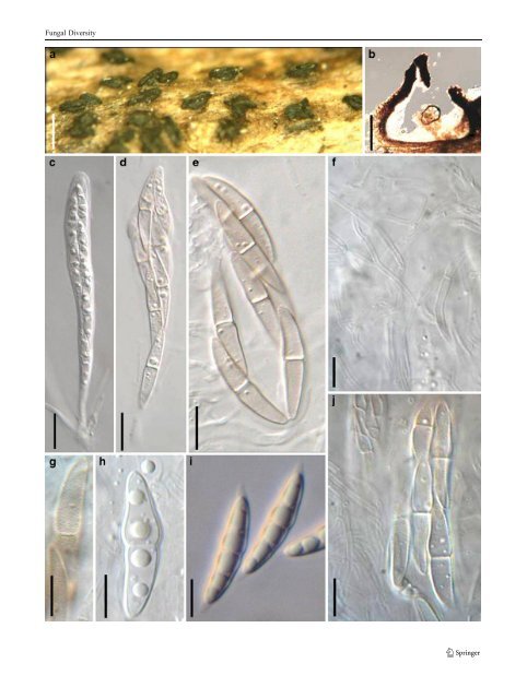
Fungal Diversity ≡ Sphaeria nucula Fr., Syst. mycol. (Lundae) 2: 466 (1823). Ascomata 200–240 μm high×200–280 μm diam., scattered, erumpent to nearly superficial, with basal wall remaining immersed in host tissue, globose to subglobose, often laterally flattened, with a flattened base not easily removed from the substrate, black, roughened; with a cylindrical or slightly compressed papilla. Papilla to 120 μm long and 150 μm high, protruding, with a pore-like ostiole (Fig. 52a). Peridium 25–30 μm wide, very thin at the base, composed of heavily pigmented pseudoparenchymatous cells near the apex, cells 2–2×6 μm diam., wall 1–3(−4) μm thick, lower sides composed of pigmented cells of textura angularis, 3–5 μm diam., wall 0.8–1.5 μm thick, ostiole wall composed of heavily pigmented and thick-walled small cells (Fig. 52b and c). Hamathecium of dense, long, septate pseudoparaphyses, 1–2 μm broad, anastomosing and branching between and above asci, embedded in mucilage (Fig. 52i). Asci 90–115× 9–11.5 μm (x ¼ 99:5 11:5mm; n=10), 8-spored, bitunicate, fissitunicate, cylindrical, with a short, narrowed, furcate pedicel which is up to 10 μm long, with a small ocular chamber (ca. 1.5μm wide×1 μm high) (J-) (Fig. 52d, e, f and h). Ascospores 17–21(−25)×(4-)5–6.5 μm (x ¼ 19:5 5:5mm, n=10), obliquely uniseriate and partially overlapping to biseriate, broadly fusoid to fusoid, with narrowly rounded ends, hyaline and lightly pigmented on very rare occasions when senescent, 1-septate, 3-septate when old, constricted at the median septum, the upper cell often broader than the lower one (Fig. 52g). Anamorph: none reported. Material examined: on decaying wood (UPS, lectotype as Sphaeria nucula Fr.). Notes Morphology Holm and Holm (1988) provided a relatively strict definition for Lophiotrema after they examined several specimens including the type materialwhichtheylectotypified. Lophiotrema was mainly defined on the unique characters of small to medium ascomata, a “Lophiotrema-type” peridium and 1-septate ascospores. In Lophiotrema, Holm and Holm (1988) considered the ascomata to be small- to medium-sized, ca. pyriform but neck often reduced, even lacking and sometimes cylindrical. The peridium was of approximately equal thickness, 20–30 μm, composed of an outer textura angularis of uniformly pigmented cells, up to 12 μm, andan inner layer of very small hyaline cells, with somewhat thickened walls. Asci are cylindrical, spores hyaline, at first 1-septate, becoming 3-septate, with distinct guttules, often with a mucilaginous sheath. Much emphasis was given to the 1-septate ascospores by Holm and Holm (1988) when they described and distinguished the three Lophiotrema species: L. boreale, L. nucula, L. vagabundum (Sacc.) Sacc. and two Fig. 51 Lophiostoma macrostomum (a–h, j from UPS, leptotype; i b from IFRD 2005). a Appearance of ascomata on the host surface. Note the raised crest-like areas and full length germ slits. b Section of the peridium. c–e Cylindro-clavate asci with ascospores arranged in a 2-3-seriate manner. f Hamathecium comprising branching and septate pseudoparaphyses. g–j Released or unreleased ascospores. Note the smooth young ascospores with terminal sheath, and the verrucose senescent ascospores. Scale bars: a=0.5 mm, b=200 μm, c–j=10 μm other unnamed species. This concept was widely accepted by later workers (Kirk et al. 2001; YuanandZhao1994). Tanaka and Harada (2003c) considered the peridium and asci to distinguish Lophiotrema from Lophiostoma, while Tang et al. (2003) introduced a new Lophiotrema species with elongated slit-like ostiole stating that the main difference between Lophiotrema and Lophiostoma were size of ascomata, structure of peridium, shape of asci and sheath of ascospores. This peridium concept however, is not supported by the lectotype specimen we examined here, which has a flattened thin-walled base. Thus the “Lophiotrema-like peridium” sensu Holm and Holm (1988) should not serve as a diagnostic character of Lophiotrema, while the ostiole, asci and ascospores might have some phylogenetic significance (Zhang et al. 2009b). No anamorph is yet known for Lophiotrema. Although the ascospores was reported by Holm and Holm (1988) to be verruculose this could not be observed in the lectotype examined under light microscope (1000 ×) in the present study. Phylogenetic study In the phylogenetic study of Lophiostoma, Massarina and related genera (Zhang et al. 2009b), Lophiotrema nucula formed a consistent and robust clade with three other Lophiotrema species: L. lignicola Yin. Zhang, J. Fourn. & K.D. Hyde, L. brunneosporum Yin. Zhang, J. Fourn. & K.D. Hyde and L. vagabundum, separate from other members of Lophiostoma and Massarina sensu stricto. This clade might represent Lophiotrema sensu stricto, however, the correctness of strains of L. vagabundum (<strong>CBS</strong> 628.86) and L. nucula (<strong>CBS</strong> 627.86) used in the phylogenetic study are not verified and warrant further study. Concluding remarks Holm and Holm (1988) distinguished Lophiostoma from Lophiotrema based on the smaller ascomata, 1-septate versus multi-septate ascospores, and peridial wall structure. However, we doubt that these distinguishing characters (size of ascomata, number of septa of ascospores) can be confidently used to separate these genera and we could not establish any characters that could reliably distinguish between these two genera. The molecular data, however, does separate Lophiostoma macrostomum and Lophiotrema nucula into separate clades and provides some support that these are separate genera. Although the strain of L. nucula
Fungal Diversity
- Page 1 and 2:
Fungal Diversity DOI 10.1007/s13225
- Page 3 and 4:
Fungal Diversity Table 1 Major circ
- Page 5 and 6:
Fungal Diversity
- Page 7 and 8:
Fungal Diversity biocontrol agent o
- Page 9 and 10:
Fungal Diversity substrates and man
- Page 11 and 12:
Fungal Diversity 2. To investigate
- Page 13 and 14:
Fungal Diversity Table 3 (continued
- Page 15 and 16:
Fungal Diversity Table 3 (continued
- Page 17 and 18:
Fungal Diversity Table 3 (continued
- Page 19 and 20:
Fungal Diversity
- Page 21 and 22:
Fungal Diversity Fig. 2 Aigialus gr
- Page 23 and 24:
Fungal Diversity Fig. 3 Amniculicol
- Page 25 and 26:
Fungal Diversity Literature: Berkel
- Page 27 and 28:
Fungal Diversity Ascorhombispora L.
- Page 29 and 30:
Fungal Diversity
- Page 31 and 32:
Fungal Diversity Fig. 8 Astrosphaer
- Page 33 and 34:
Fungal Diversity Fig. 9 Asymmetrico
- Page 35 and 36:
Fungal Diversity Notes Morphology B
- Page 37 and 38:
Fungal Diversity Generic descriptio
- Page 39 and 40:
Fungal Diversity Anamorph: none rep
- Page 41 and 42:
Fungal Diversity Fig. 14 Bimuria no
- Page 43 and 44:
Fungal Diversity Fig. 15 Bricookea
- Page 45 and 46:
Fungal Diversity Fig. 16 Byssolophi
- Page 47 and 48:
Fungal Diversity Notes Morphology B
- Page 49 and 50:
Fungal Diversity the reaction of pe
- Page 51 and 52:
Fungal Diversity
- Page 53 and 54: Fungal Diversity Fig. 21 Chaetomast
- Page 55 and 56: Fungal Diversity
- Page 57 and 58: Fungal Diversity Fig. 23 Cilioplea
- Page 59 and 60: Fungal Diversity with one or two ve
- Page 61 and 62: Fungal Diversity Moreau 1953; Munk
- Page 63 and 64: Fungal Diversity Material examined:
- Page 65 and 66: Fungal Diversity Fig. 28 Dothidotth
- Page 67 and 68: Fungal Diversity Fig. 29 Dubitatio
- Page 69 and 70: Fungal Diversity assigned Entodesmi
- Page 71 and 72: Fungal Diversity fusoid to somewhat
- Page 73 and 74: Fungal Diversity Fig. 33 Hadrospora
- Page 75 and 76: Fungal Diversity Fig. 34 Halotthia
- Page 77 and 78: Fungal Diversity Notes Morphology H
- Page 79 and 80: Fungal Diversity some effused Hypox
- Page 81 and 82: Fungal Diversity Fig. 38 Isthmospor
- Page 83 and 84: Fungal Diversity Fig. 39 Kalmusia e
- Page 85 and 86: Fungal Diversity ascospores were br
- Page 87 and 88: Fungal Diversity furcate pedicel an
- Page 89 and 90: Fungal Diversity Anamorph: none rep
- Page 91 and 92: Fungal Diversity
- Page 93 and 94: Fungal Diversity Material examined:
- Page 95 and 96: Fungal Diversity Fig. 46 Lewia scro
- Page 97 and 98: Fungal Diversity Fig. 47 Lichenopyr
- Page 99 and 100: Fungal Diversity Loculohypoxylon M.
- Page 101 and 102: Fungal Diversity cells small heavil
- Page 103: Fungal Diversity upper place, septa
- Page 107 and 108: Fungal Diversity (CBS 627.86) was i
- Page 109 and 110: Fungal Diversity Fig. 54 Mamillisph
- Page 111 and 112: Fungal Diversity Fig. 55 Massarina
- Page 113 and 114: Fungal Diversity phaeria as a synon
- Page 115 and 116: Fungal Diversity 5-8 μm diam., ind
- Page 117 and 118: Fungal Diversity cell wall
- Page 119 and 120: Fungal Diversity Fig. 60 Mixtura sa
- Page 121 and 122: Fungal Diversity Fig. 61 Montagnula
- Page 123 and 124: Fungal Diversity spored, bitunicate
- Page 125 and 126: Fungal Diversity Fig. 64 Murispora
- Page 127 and 128: Fungal Diversity Type species Neoph
- Page 129 and 130: Fungal Diversity brown, 8-septate,
- Page 131 and 132: Fungal Diversity Fig. 68 Ohleria mo
- Page 133 and 134: Fungal Diversity Fig. 69 Ohleriella
- Page 135 and 136: Fungal Diversity Fig. 70 Ophiobolus
- Page 137 and 138: Fungal Diversity Type species Ostro
- Page 139 and 140: Fungal Diversity
- Page 141 and 142: Fungal Diversity (Shoemaker and Bab
- Page 143 and 144: Fungal Diversity ium thin, composed
- Page 145 and 146: Fungal Diversity Fig. 76 Platysporo
- Page 147 and 148: Fungal Diversity Fig. 77 1 Pleomass
- Page 149 and 150: Fungal Diversity Fig. 78 Pleophragm
- Page 151 and 152: Fungal Diversity papillate, ostiola
- Page 153 and 154: Fungal Diversity Williams 1963; Mal
- Page 155 and 156:
Fungal Diversity Generic descriptio
- Page 157 and 158:
Fungal Diversity composed of one ce
- Page 159 and 160:
Fungal Diversity Fig. 84 Saccharico
- Page 161 and 162:
Fungal Diversity and nearly black a
- Page 163 and 164:
Fungal Diversity dense, long trabec
- Page 165 and 166:
Fungal Diversity
- Page 167 and 168:
Fungal Diversity
- Page 169 and 170:
Fungal Diversity Anamorphs reported
- Page 171 and 172:
Fungal Diversity
- Page 173 and 174:
Fungal Diversity
- Page 175 and 176:
Fungal Diversity Fig. 94 Westerdyke
- Page 177 and 178:
Fungal Diversity Fig. 95 Wettsteini
- Page 179 and 180:
Fungal Diversity Fig. 96 Wilmia bra
- Page 181 and 182:
Fungal Diversity Current name: Astr
- Page 183 and 184:
Fungal Diversity spores are actuall
- Page 185 and 186:
Fungal Diversity Fig. 100 Sporormie
- Page 187 and 188:
Fungal Diversity
- Page 189 and 190:
Fungal Diversity Fig. 102 Kriegerie
- Page 191 and 192:
Fungal Diversity Phylogenetic study
- Page 193 and 194:
Fungal Diversity Fig. 104 Zeuctomor
- Page 195 and 196:
Fungal Diversity Fig. 105 Muroia ni
- Page 197 and 198:
Fungal Diversity pseudoparenchymato
- Page 199 and 200:
Fungal Diversity Eremodothis Arx, K
- Page 201 and 202:
Fungal Diversity Type species: Macr
- Page 203 and 204:
Fungal Diversity ascospores of Plat
- Page 205 and 206:
Fungal Diversity monoceras Alcorn n
- Page 207 and 208:
Fungal Diversity tomataceae, Melano
- Page 209 and 210:
Fungal Diversity Table 4 (continued
- Page 211 and 212:
Fungal Diversity 1987b). Based on a
- Page 213 and 214:
Fungal Diversity only do so under v
- Page 215 and 216:
Fungal Diversity Dennis RWG (1968)
- Page 217 and 218:
Fungal Diversity Kirk PM, Cannon PF
- Page 219 and 220:
Fungal Diversity Saccardo PA (1880)
- Page 221:
Fungal Diversity Winter G (1887) As