Fungal Diversity pedicel which is up to 45 μm long, and a low ocular chamber (ca. 2μm wide×1 μm high) (Fig. 39d, e and f). Ascospores 15–18×5.5–6.5 μm (x ¼ 16:3 5:8mm, n=10), biseriate, narrowly ovoid to clavate, pale brown, 3-distoseptate, without constriction, smooth-walled (Fig. 39g, h and i). Anamorph: none reported. Material examined: BELGIUM, Dolembreux, on branchlets and pieces of stumps of Sarothamnus scoparius from woodland, Oct. 1922, V. Mouton (BR 101525–63, holotype). Notes Morphology Kalmusia was formally established by von Niessl (1872), and is mainly characterized as “immersed, sphaeroid ascoma with central, stout papilla, surrounded by hyphae in the substrate, stipitate asci with septate pseudoparaphyses, and brown, 3-septate, inequilateral ascospores” (Barr 1992a). The most morphologically comparable genus to Kalmusia is Thyridaria, which had been treated as a subgenus under Kalmusia (Lindau 1897), and was subsequently transferred to Platystomaceae in Melanommatales (Barr 1987b, 1990a). Compared to Thyridaria, Kalmusia has sphaeroid ascomata, a peridium of small pseudoparenchymatous cells, basal asci and very thin pseudoparaphyses, thus it was assigned to Phaeosphaeriaceae of the <strong>Pleosporales</strong> by Barr (1990a), and the genus is utilized to accommodate both K. ebuli and K. clivensis (Berk. & Broome) M.E. Barr, as well as closely related species, i.e. K. utahensis (Ellis & Everh.) Huhndorf & M.E. Barr and K. coniothyrium (Fuckel) Huhndorf (Barr 1992a). But this proposal is questionable, as the clavate, distoseptate ascospores, as well as the clavate asci with very long pedicels are uncommon in Phaeosphaeriaceae, and most recent phylogenetic study indicated that some species of Kalmusia reside outside of Phaeosphaeriaceae (Zhang et al. 2009a). Phylogenetic study Both Kalmusia scabrispora Teng Kaz. Tanaka, Y. Harada & M.E. Barr and K. brevispora (Nagas. & Y. Otani) Yin. Zhang, Kaz. Tanaka & C.L. Schoch reside in the clade of Montagnulaceae (Zhang et al. 2009a). Familial placement of Kalmusia can only be verified after the DNA sequences of the generic type (K. ebuli) are obtained. Concluding remarks Kalmusia is distinct amongst the <strong>Pleosporales</strong> as it has pale brown ascospores with indistinct distosepta and clavate asci with long pedicels. Although both K. scabrispora and K. brevispora reside in the clade of Montagnulaceae, they both lack the distoseptate ascospores that are possessed by the generic type (K. ebuli). Thus, the familial placement of Kalmusia is still undetermined. Karstenula Speg., Decades Mycologicae Italicae ad no. 94 (in sched.) (1879). (Montagnulaceae) Generic description Habitat terrestrial, saprobic. Ascomata rarely small-, usually medium-sized, immersed usually under thin clypeus, scattered to gregarious, with flattened top and rounded pore-like ostiole, coriaceous. Peridium 2-layered, outer layer composed of reddish brown to dark brown small cells, inner layer of pale compressed cells. Hamathecium of dense, cellular pseudoparaphyses. Asci cylindrical to cylindro-clavate with short furcate pedicel. Ascospores muriform, ellipsoid to fusoid, reddish brown to dark brown. Anamorphs reported for the genus: Microdiplodia (Constantinescu 1993). Literature: Barr 1990a; Eriksson and Hawksworth 1991; Kodsueb et al. 2006a; Munk 1957; Zhang et al. 2009a. Type species Karstenula rhodostoma (Alb. & Schwein.) Speg., Decades Mycologicae Italicae no. 94. (1879). (Fig. 40) ≡ Sphaeria rhodostoma Alb. & Schwein., Consp. fung. (Leipzig): 43 (1805). Ascomata 250–430 μm high×450–650 μm diam., scattered or gregarious, immersed in the subiculum which sometimes sloths off, globose or subglobose, black, flattened top often white or reddish and sometimes slightly protruding out of the substrate surface, usually with a wide opening ostiole after removing the cover, coriaceous (Fig. 40a and b). Peridium 30–40 μm wide, comprising two cell types, outer region 1- layered, composed of relatively small heavily pigmented thick-walled compressed cells, cells 2–4×5–10 μm diam., cell wall 2–4 μm thick, inner layer cells larger and wall thinner, comprising cells of textura angularis, merging with pseudoparaphyses (Fig. 40c and d). Hamathecium of dense, long cellular pseudoparaphyses 2–3.5 μm broad, septate, branching or anastomosing not observed. Asci 150–210× 12.5–15 μm (x ¼ 182 13:1mm, n=10), 8-spored, bitunicate, fissitunicate, cylindrical, with a broad, furcate pedicel which is 12–35 μm long, and with an ocular chamber (to 4 μm wide× 3 μm high) (Fig. 40e and f). Ascospores 20–26×7.5–10 μm (x ¼ 22:4 8mm, n=10), obliquely uniseriate and partially overlapping, ellipsoid, reddish brown, with 3 transverse septa and a vertical septum in one or two central cells, constricted at the septa, verruculose (Fig. 40g, h and i). Anamorph: Microdiplodia frangulae Allesch. (Constantinescu 1993). Conidiomata globose to subglobose, 330–495 μm diam., in subiculum. Conidia 9–13×4–5 μm, reddish brown, 1- septate (information obtained from Barr 1990a).
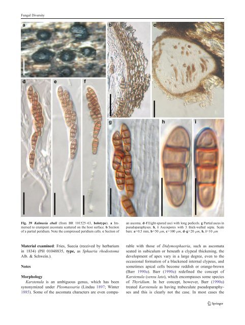
Fungal Diversity Fig. 39 Kalmusia ebuli (from BR 101525–63, holotype). a Immersed to erumpent ascomata scattered on the host surface. b Section of a partial peridium. Note the compressed peridium cells. c Section of an ascoma. d–f Eight-spored asci with long pedicels. g Partial ascus in pseudoparaphyses. h, i Ascospores with 3 thick-walled septa. Scale bars: a=0.5 mm, b=50 μm, c=100 μm, d–g=20 μm, h, i=10 μm Material examined: Fries, Suecia (received by herbarium in 1834) (PH 01048835, type, as Sphaeria rhodostoma Alb. & Schwein.). Notes Morphology Karstenula is an ambiguous genus, which has been synonymized under Pleomassaria (Lindau 1897; Winter 1885). Some of the ascomata characters are even comparable with those of Didymosphaeria, such as ascomata seated in subiculum or beneath a clypeal thickening, the development of apex vary in a large degree, even to the occasional formation of a blackened internal clypeus, and sometimes apical cells become reddish or orange-brown (Barr 1990a). Barr (1990a) redefined the concept of Karstenula (sensu lato), which encompasses some species of Thyridium. In her concept, however, Barr (1990a) treated Karstenula as having trabeculate pseudoparaphyses and this is clearly not the case. In most cases the
- Page 1 and 2:
Fungal Diversity DOI 10.1007/s13225
- Page 3 and 4:
Fungal Diversity Table 1 Major circ
- Page 5 and 6:
Fungal Diversity
- Page 7 and 8:
Fungal Diversity biocontrol agent o
- Page 9 and 10:
Fungal Diversity substrates and man
- Page 11 and 12:
Fungal Diversity 2. To investigate
- Page 13 and 14:
Fungal Diversity Table 3 (continued
- Page 15 and 16:
Fungal Diversity Table 3 (continued
- Page 17 and 18:
Fungal Diversity Table 3 (continued
- Page 19 and 20:
Fungal Diversity
- Page 21 and 22:
Fungal Diversity Fig. 2 Aigialus gr
- Page 23 and 24:
Fungal Diversity Fig. 3 Amniculicol
- Page 25 and 26:
Fungal Diversity Literature: Berkel
- Page 27 and 28:
Fungal Diversity Ascorhombispora L.
- Page 29 and 30:
Fungal Diversity
- Page 31 and 32: Fungal Diversity Fig. 8 Astrosphaer
- Page 33 and 34: Fungal Diversity Fig. 9 Asymmetrico
- Page 35 and 36: Fungal Diversity Notes Morphology B
- Page 37 and 38: Fungal Diversity Generic descriptio
- Page 39 and 40: Fungal Diversity Anamorph: none rep
- Page 41 and 42: Fungal Diversity Fig. 14 Bimuria no
- Page 43 and 44: Fungal Diversity Fig. 15 Bricookea
- Page 45 and 46: Fungal Diversity Fig. 16 Byssolophi
- Page 47 and 48: Fungal Diversity Notes Morphology B
- Page 49 and 50: Fungal Diversity the reaction of pe
- Page 51 and 52: Fungal Diversity
- Page 53 and 54: Fungal Diversity Fig. 21 Chaetomast
- Page 55 and 56: Fungal Diversity
- Page 57 and 58: Fungal Diversity Fig. 23 Cilioplea
- Page 59 and 60: Fungal Diversity with one or two ve
- Page 61 and 62: Fungal Diversity Moreau 1953; Munk
- Page 63 and 64: Fungal Diversity Material examined:
- Page 65 and 66: Fungal Diversity Fig. 28 Dothidotth
- Page 67 and 68: Fungal Diversity Fig. 29 Dubitatio
- Page 69 and 70: Fungal Diversity assigned Entodesmi
- Page 71 and 72: Fungal Diversity fusoid to somewhat
- Page 73 and 74: Fungal Diversity Fig. 33 Hadrospora
- Page 75 and 76: Fungal Diversity Fig. 34 Halotthia
- Page 77 and 78: Fungal Diversity Notes Morphology H
- Page 79 and 80: Fungal Diversity some effused Hypox
- Page 81: Fungal Diversity Fig. 38 Isthmospor
- Page 85 and 86: Fungal Diversity ascospores were br
- Page 87 and 88: Fungal Diversity furcate pedicel an
- Page 89 and 90: Fungal Diversity Anamorph: none rep
- Page 91 and 92: Fungal Diversity
- Page 93 and 94: Fungal Diversity Material examined:
- Page 95 and 96: Fungal Diversity Fig. 46 Lewia scro
- Page 97 and 98: Fungal Diversity Fig. 47 Lichenopyr
- Page 99 and 100: Fungal Diversity Loculohypoxylon M.
- Page 101 and 102: Fungal Diversity cells small heavil
- Page 103 and 104: Fungal Diversity upper place, septa
- Page 105 and 106: Fungal Diversity
- Page 107 and 108: Fungal Diversity (CBS 627.86) was i
- Page 109 and 110: Fungal Diversity Fig. 54 Mamillisph
- Page 111 and 112: Fungal Diversity Fig. 55 Massarina
- Page 113 and 114: Fungal Diversity phaeria as a synon
- Page 115 and 116: Fungal Diversity 5-8 μm diam., ind
- Page 117 and 118: Fungal Diversity cell wall
- Page 119 and 120: Fungal Diversity Fig. 60 Mixtura sa
- Page 121 and 122: Fungal Diversity Fig. 61 Montagnula
- Page 123 and 124: Fungal Diversity spored, bitunicate
- Page 125 and 126: Fungal Diversity Fig. 64 Murispora
- Page 127 and 128: Fungal Diversity Type species Neoph
- Page 129 and 130: Fungal Diversity brown, 8-septate,
- Page 131 and 132: Fungal Diversity Fig. 68 Ohleria mo
- Page 133 and 134:
Fungal Diversity Fig. 69 Ohleriella
- Page 135 and 136:
Fungal Diversity Fig. 70 Ophiobolus
- Page 137 and 138:
Fungal Diversity Type species Ostro
- Page 139 and 140:
Fungal Diversity
- Page 141 and 142:
Fungal Diversity (Shoemaker and Bab
- Page 143 and 144:
Fungal Diversity ium thin, composed
- Page 145 and 146:
Fungal Diversity Fig. 76 Platysporo
- Page 147 and 148:
Fungal Diversity Fig. 77 1 Pleomass
- Page 149 and 150:
Fungal Diversity Fig. 78 Pleophragm
- Page 151 and 152:
Fungal Diversity papillate, ostiola
- Page 153 and 154:
Fungal Diversity Williams 1963; Mal
- Page 155 and 156:
Fungal Diversity Generic descriptio
- Page 157 and 158:
Fungal Diversity composed of one ce
- Page 159 and 160:
Fungal Diversity Fig. 84 Saccharico
- Page 161 and 162:
Fungal Diversity and nearly black a
- Page 163 and 164:
Fungal Diversity dense, long trabec
- Page 165 and 166:
Fungal Diversity
- Page 167 and 168:
Fungal Diversity
- Page 169 and 170:
Fungal Diversity Anamorphs reported
- Page 171 and 172:
Fungal Diversity
- Page 173 and 174:
Fungal Diversity
- Page 175 and 176:
Fungal Diversity Fig. 94 Westerdyke
- Page 177 and 178:
Fungal Diversity Fig. 95 Wettsteini
- Page 179 and 180:
Fungal Diversity Fig. 96 Wilmia bra
- Page 181 and 182:
Fungal Diversity Current name: Astr
- Page 183 and 184:
Fungal Diversity spores are actuall
- Page 185 and 186:
Fungal Diversity Fig. 100 Sporormie
- Page 187 and 188:
Fungal Diversity
- Page 189 and 190:
Fungal Diversity Fig. 102 Kriegerie
- Page 191 and 192:
Fungal Diversity Phylogenetic study
- Page 193 and 194:
Fungal Diversity Fig. 104 Zeuctomor
- Page 195 and 196:
Fungal Diversity Fig. 105 Muroia ni
- Page 197 and 198:
Fungal Diversity pseudoparenchymato
- Page 199 and 200:
Fungal Diversity Eremodothis Arx, K
- Page 201 and 202:
Fungal Diversity Type species: Macr
- Page 203 and 204:
Fungal Diversity ascospores of Plat
- Page 205 and 206:
Fungal Diversity monoceras Alcorn n
- Page 207 and 208:
Fungal Diversity tomataceae, Melano
- Page 209 and 210:
Fungal Diversity Table 4 (continued
- Page 211 and 212:
Fungal Diversity 1987b). Based on a
- Page 213 and 214:
Fungal Diversity only do so under v
- Page 215 and 216:
Fungal Diversity Dennis RWG (1968)
- Page 217 and 218:
Fungal Diversity Kirk PM, Cannon PF
- Page 219 and 220:
Fungal Diversity Saccardo PA (1880)
- Page 221:
Fungal Diversity Winter G (1887) As