

Pleosporales - CBS - KNAW
Pleosporales - CBS - KNAW
Pleosporales - CBS - KNAW

You also want an ePaper? Increase the reach of your titles
YUMPU automatically turns print PDFs into web optimized ePapers that Google loves.
Fungal Diversity<br />
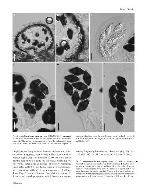
Fig. 6 Ascorhombispora aquatica (from HKU(M) 10859, holotype).<br />
a Section of an ascoma. b Section of a partial peridium. c Immature<br />
ascus. d–f Mature asci with ascospores. Note the deliquescent ascal<br />
wall in f. Note the wide, dark band in the medium septum of<br />
ascospores in d and e and the mucilaginous sheath and paler end cells<br />
in e and f. Scale bars: a=20 μm, b–f=15 μm (figures referred to Cai<br />
and Hyde 2007)<br />
subglobose, not easily removed from the substrate, wall black,<br />
coriaceous, roughened, apex usually widely porate, with or<br />
without papilla (Fig. 7a). Peridium 70–90 μm wide, thicker<br />
near the base where it is up to 180 μm wide, comprising two<br />
cell types, outer cells composed of heavily pigmented<br />
small cells, cells 3–5 μm diam., inner layer composed of<br />
less pigmented cells of textura angularis, 10–20 μm<br />
diam. (Fig. 7b and c). Hamathecium of dense, septate, 2–<br />
3 μm broad, pseudoparaphyses which branch and anastomosing<br />
frequently between and above asci (Fig. 7d). Asci<br />
(180-)200–280×28–43 μm (x ¼ 230 35mm, n=10), 8-<br />
Fig. 7 Asteromassaria macrospora (from L, 1004). a Ascomata<br />
clustered in a group breaking through the host surface. b Section of an<br />
ascoma. c Section of a partial peridium. Note the cells of textura<br />
angularis. d Pseudoparaphyses. Note the branches. e Upper part of the<br />
ascus illustrating the ocular chamber. f Ascus with a short pedicel. g–j<br />
Ascospores. Note the mucilaginous sheath in G and minutely verruculose<br />
ornamentation in J. Scale bars: a=0.5 mm, b, c=100 μm, d–j=10 μm<br />
b


