Dernière édition Attention: Le pdf pèse environ 17 - BFH-TI - Berner ...
Dernière édition Attention: Le pdf pèse environ 17 - BFH-TI - Berner ...
Dernière édition Attention: Le pdf pèse environ 17 - BFH-TI - Berner ...
- Keine Tags gefunden...

Erfolgreiche ePaper selbst erstellen
Machen Sie aus Ihren PDF Publikationen ein blätterbares Flipbook mit unserer einzigartigen Google optimierten e-Paper Software.
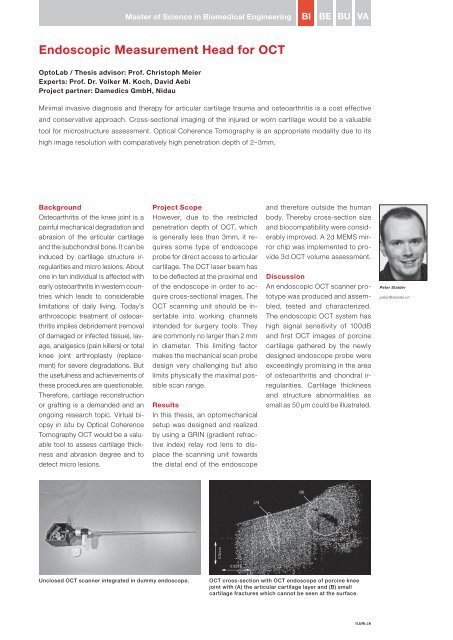
Master of Science in Biomedical EngineeringBIBEBUVAEndoscopic Measurement Head for OCTOptoLab / Thesis advisor: Prof. Christoph MeierExperts: Prof. Dr. Volker M. Koch, David AebiProject partner: Damedics GmbH, NidauMinimal invasive diagnosis and therapy for articular cartilage trauma and osteoarthritis is a cost effectiveand conservative approach. Cross-sectional imaging of the injured or worn cartilage would be a valuabletool for microstructure assessment. Optical Coherence Tomography is an appropriate modality due to itshigh image resolution with comparatively high penetration depth of 2–3mm.BackgroundOsteoarthritis of the knee joint is apainful mechanical degradation andabrasion of the articular cartilageand the subchondral bone. It can beinduced by cartilage structure irregularitiesand micro lesions. Aboutone in ten individual is affected withearly osteoarthritis in western countrieswhich leads to considerablelimitations of daily living. Today’sarthroscopic treatment of osteoarthritisimplies debridement (removalof damaged or infected tissue), lavage,analgesics (pain killers) or totalknee joint arthroplasty (replacement)for severe degradations. Butthe usefulness and achievements ofthese procedures are questionable.Therefore, cartilage reconstructionor grafting is a demanded and anongoing research topic. Virtual biopsyin situ by Optical CoherenceTomography OCT would be a valuabletool to assess cartilage thicknessand abrasion degree and todetect micro lesions.Project ScopeHowever, due to the restrictedpenetration depth of OCT, whichis generally less than 3mm, it requiressome type of endoscopeprobe for direct access to articularcartilage. The OCT laser beam hasto be deflected at the proximal endof the endoscope in order to acquirecross-sectional images. TheOCT scanning unit should be insertableinto working channelsintended for surgery tools. Theyare commonly no larger than 2 mmin diameter. This limiting factormakes the mechanical scan probedesign very challenging but alsolimits physically the maximal possiblescan range.ResultsIn this thesis, an optomechanicalsetup was designed and realizedby using a GRIN (gradient refractiveindex) relay rod lens to displacethe scanning unit towardsthe distal end of the endoscopeand therefore outside the humanbody. Thereby cross-section sizeand biocompatibility were considerablyimproved. A 2d MEMS mirrorchip was implemented to provide3d OCT volume assessment.DiscussionAn endoscopic OCT scanner prototypewas produced and assembled,tested and characterized.The endoscopic OCT system hashigh signal sensitivity of 100dBand first OCT images of porcinecartilage gathered by the newlydesigned endoscope probe wereexceedingly promising in the areaof osteoarthritis and chondral irregularities.Cartilage thicknessand structure abnormalities assmall as 50 μm could be illustrated.Peter Stalderpeter@stalder.chUnclosed OCT scanner integrated in dummy endoscope.OCT cross-section with OCT endoscope of porcine kneejoint with (A) the articular cartilage layer and (B) smallcartilage fractures which cannot be seen at the surface.ti.bfh.ch49