- Page 2:
A..2^ BOUGHT WITH THE INCOME FROM T
- Page 5:
Tracing from nature of a mesial lin
- Page 13 and 14:
TABLE OF CONTENTS VOLUME ONE PREFAC
- Page 15 and 16:
§ 36. The Travelling Organs in Rel
- Page 17 and 18:
CONTENTS xi Rhythmic Movements—An
- Page 19 and 20:
CONTENTS xiii THE MOVEMENTS AND FUN
- Page 21 and 22:
§§ . CONTENTS XV INTELLIGENCE OF
- Page 23 and 24:
CONTENTS xvii THE OSSEOUS AND MUSCU
- Page 25 and 26:
§ 420. The Flight of Birds divisib
- Page 27 and 28:
PREFACE The present work has attain
- Page 29 and 30:
INTRODUCTION The present work natur
- Page 31 and 32:
INTRODUCTION xxv cules being so arr
- Page 33 and 34:
INTRODUCTION xxvii is the ancient w
- Page 35 and 36:
INTRODUCTION xxix ments met with in
- Page 37 and 38:
INTRODUCTION xxxi organisms there i
- Page 39 and 40:
DESIGN IN NATURE INORGANIC AND ORGA
- Page 41 and 42:
STRAIGHT-LINE AND OTHER FORMATIONS
- Page 43 and 44:
ARRANGEMENTS COMMON TO CRYSTALS, PL
- Page 45 and 46:
ARRANGEMENTS COMMON TO CRYSTALS, PL
- Page 47 and 48:
ARRANGEMENTS COMMON TO CRYSTALS, PL
- Page 49 and 50:
PREVALENCE OF SPIRAL ARRANGEMENTS 1
- Page 51 and 52:
PREVALENCE OF SPIRAL ARRANGEMENTS P
- Page 53 and 54:
PREVALENCE OF SPIRAL ARRANGEMENTS P
- Page 55 and 56:
VOL. I PREVALENCE OF SPIRAL ARRANGE
- Page 57 and 58:
ORIGIN OF SPIRAL STRUCTURES 19 and
- Page 59 and 60:
SPIRAL ARRANGEMENTS IN PLANTS 21 |3
- Page 61 and 62:
SPIRAL ARRANGEMENTS IN PLANTS 23 PL
- Page 63 and 64:
SPIRAL ARRANGEMENTS IN PLANTS PLATE
- Page 65 and 66:
Fig. 3. SPIRAL ARR'ANGEMENTS IN ANI
- Page 67 and 68:
SPIRAL ARRANGEMENTS IN ANIMALS 29 P
- Page 69 and 70:
SPIRAL ARRANGEMENTS IN ANIMALS PLAT
- Page 71 and 72:
SPIRAL ARRANGEMENTS IN ANIMALS 33 P
- Page 73 and 74:
SPIRAL ARRANGEMENTS IN ANIMALS Fir.
- Page 75 and 76:
CBERJtAU, SPIRAL ARRANGEMENTS IN AN
- Page 77 and 78:
RADIATING AND CONCENTRIC ARRANGEMEN
- Page 79 and 80:
RADIATING AND CONCENTRIC ARRANGEMEN
- Page 81 and 82:
RADIATING AND CONCENTRIC ARRANGEMEN
- Page 83 and 84:
1''IG. 1. RADIATING AND CONCENTRIC
- Page 85 and 86:
§ 9. Dendritic or Branching Moveme
- Page 87 and 88:
VOL. I, DENDRITIC OR BRANCHING ARRA
- Page 89 and 90:
DENDRITIC OR BRANCHING ARRANGEMENTS
- Page 91 and 92:
DENDRITIC OR BRANCHING ARRANGEMENTS
- Page 93 and 94:
DENDRITIC OR BRANCHING ARRANGEMENTS
- Page 95 and 96:
VOL. I. DENDRITIC OR BRANCHING ARRA
- Page 97 and 98:
BRANCHING AND RADIATING ARRANGEMENT
- Page 99 and 100:
BRANCHING AND RADIATING ARRANGEMENT
- Page 101 and 102:
HEXAGONAL STRUCTURES AND SEGMENTATI
- Page 103 and 104:
HEXAGONAL STRUCTURES 65 Fig. 20.—
- Page 105 and 106:
CLEAVAGE AND SEGMENTATION 67 PLATE
- Page 107 and 108:
LONGITUDINAL AND TRANSVERSE CLEAVAG
- Page 109 and 110:
CONVOLUTIONS IN HARD AND SOFT PARTS
- Page 111 and 112:
RADIATING AND BRANCHING ARRANGEMENT
- Page 113 and 114:
LONGITUDINAL AND TRANSVERSE CLEAVAG
- Page 115 and 116:
LONGITUDINAL, RADIATING, AND TRANSV
- Page 117 and 118:
LONGITUDINAL, RADIATING, AND TRANSV
- Page 119 and 120:
VOL. I. LONGITUDINAL AND RADIATING
- Page 121 and 122:
LONGITUDINAL, RADIATING, AND TRANSV
- Page 123 and 124:
TRAVELLING ORGANS FOR LAND, WATER,
- Page 125 and 126:
j^-«i Fig, S. FiG. Fig. 10. FIGURE
- Page 127 and 128:
vol.. I, RECAPITULATION 89 PLATE LI
- Page 129 and 130:
RECAPITULATION 91 The same thing, w
- Page 131 and 132:
ATOMS AND MOLECULES 93 some anthrop
- Page 133 and 134:
CONSERVATION OF ENERGY 95 secretion
- Page 135 and 136:
THE REPRODUCTIVE ELEMENTS OF PLANTS
- Page 137 and 138:
SPIRAL STRUCTURES AND MOVEMENTS IN
- Page 139 and 140:
ATOMS AND MOLECULES IN DEAD AND LIV
- Page 141 and 142: UNITY OF PLAN IN NATURE 103 arrange
- Page 143 and 144: force is so feeble that the chain o
- Page 145 and 146: FIG. nG.4 FIG. 5 m^0'^i^ FIG. 6 FIG
- Page 147 and 148: •'•'•;.v':^Hv'i4t^ ''•',>;'
- Page 149 and 150: THE BAR-MAGNET iii number as polar
- Page 151 and 152: THE COMPASS 113 of gelatine and pla
- Page 153 and 154: MAGNETISM, ELECTRICITY, LIGHT, HEAT
- Page 155 and 156: MAGNETISM, ELECTRICITY, LIGHT, HEAT
- Page 157 and 158: ATMOSPHERIC AND OTHER ELECTRICITY 1
- Page 159 and 160: ATMOSPHERIC AND OTHER ELECTRICITY 1
- Page 161 and 162: ANIMAL MAGNETISM 123 the electric o
- Page 163 and 164: LINES OF COMMUNICATION AND FORCE 12
- Page 165 and 166: LINES OF COMMUNICATION AND FORCE 12
- Page 167 and 168: LINES OF COMMUNICATION AND FORCE 12
- Page 169 and 170: LINES OF COMMUNICATION AND FORCE 13
- Page 171 and 172: A BEGINNINGS OF NERVOUS SYSTEM Fig.
- Page 173 and 174: BEGINNINGS OF NERVOUS SYSTEM 135 So
- Page 175 and 176: VOL I. ATOMS, MOLECULES, AND CELLS
- Page 177 and 178: ATOMS, MOLECULES, AND CELLS AS FACT
- Page 179 and 180: ATOMS, MOLECULES, AND CELLS AS FACT
- Page 181 and 182: a' ATOMS, MOLECULES, AND CELLS AS F
- Page 183 and 184: ATOMS, MOLECULES, AND CELLS AS FACT
- Page 185 and 186: EVIDENCES OF DESIGN IN REPRODUCTIVE
- Page 187 and 188: EVIDENCES OF DESIGN IN. REPRODUCTIV
- Page 189 and 190: EVIDENCES OF DESIGN IN REPRODUCTIVE
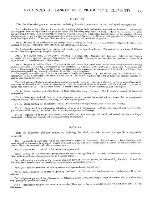
- Page 191: EVIDENCES OF DESIGN IN REPRODUCTIVE
- Page 195 and 196: EVIDENCES OF DESIGN IN REPRODUCTIVE
- Page 197 and 198: EVIDENCES OF DESIGN IN REPRODUCTIVE
- Page 199 and 200: ADVANCE IN LOWER PLANT AND ANIMAL F
- Page 201 and 202: ADVANCE IN LOWER PLANT AND ANIMAL F
- Page 203 and 204: ADVANCE IN LOWER PLANT AND ANIMAL F
- Page 205 and 206: ADVANCE IN LOWER PLANT AND ANIMAL F
- Page 207 and 208: ADVANCE IN LOWER PLANT AND ANIMAL F
- Page 209 and 210: ADVANCE IN LOWER PLANT AND ANIMAL F
- Page 211 and 212: ADVANCE IN LOWER PLANT AND ANIMAL F
- Page 213 and 214: ADVANCE IN LOWER PLANT AND ANIMAL F
- Page 215 and 216: ADVANCE IN LOWER PLANT AND ANIMAL F
- Page 217 and 218: ADVANCE IN LOWER PLANT AND ANIMAL F
- Page 219 and 220: THE VISIBLE AND INVISIBLE WORLD i8i
- Page 221 and 222: THE VISIBLE AND INVISIBLE WORLD 183
- Page 223 and 224: NEW THEORY OF MATTER 185 " To obtai
- Page 225 and 226: NEW THEORY OF MATTER 187 in action
- Page 227 and 228: NEW THEORY OF MATTER 189 the outset
- Page 229 and 230: INTERACTION BETWEEN MENTAL AND MATE
- Page 231 and 232: MATTER AND FORCE IN INORGANIC AND O
- Page 233 and 234: Molybdenum THE ELEMENTS AND THEIR C
- Page 235 and 236: HAECKEL'S BELIEF IN THE OMNIPOTENCE
- Page 237 and 238: HAECKEL'S BELIEF IN THE OMNIPOTENCE
- Page 239 and 240: MECHANICAL VIEWS OF KANT AND LAPLAC
- Page 241 and 242: PROFESSOR HUXLEY'S VIEWS ON EVOLUTI
- Page 243 and 244:
PROFESSOR HUXLEY'S VIEWS ON EVOLUTI
- Page 245 and 246:
PROFESSOR HUXLEY'S VIEWS ON EVOLUTI
- Page 247 and 248:
PROFESSOR HUXLEY'S VIEWS ON EVOLUTI
- Page 249 and 250:
PROFESSOR HUXLEY'S VIEWS ON EVOLUTI
- Page 251 and 252:
PROFESSOR HUXLEY'S VIEWS ON EVOLUTI
- Page 253 and 254:
PROFESSOR HUXLEY'S VIEWS ON EVOLUTI
- Page 255 and 256:
THE TRAVELLING ORGANS OF ANIMALS 21
- Page 257 and 258:
THE TRAVELLING ORGANS OF ANIMALS 21
- Page 259 and 260:
TRAVELLING ORGANS IN RELATION TO EN
- Page 261 and 262:
KANT'S AND SPENCER'S VIEWS OF MATTE
- Page 263 and 264:
KANT'S AND SPENCER'S VIEWS OF MATTE
- Page 265 and 266:
SCRIPTURAL ACCOUNT OF CREATION 227
- Page 267 and 268:
GEOLOGY AS BEARING ON CREATION 229
- Page 269 and 270:
SIMPLE AND COMPLEX PLANTS AND ANIMA
- Page 271 and 272:
PLANTS AND ANIMALS IMPROVABLE UP TO
- Page 273 and 274:
EVERYTHING CONTROLLED AND UNDER SUP
- Page 275 and 276:
THE UNIVERSE AS A WORKING SYSTEM 23
- Page 277 and 278:
THE UNIVERSE AS A WORKING SYSTEM 23
- Page 279 and 280:
ENVIRONMENT 241 To take other examp
- Page 281 and 282:
INSTINCT 243 advance in plants and
- Page 283 and 284:
INSTINCT AND INTELLIGENCE 245 custo
- Page 285 and 286:
EFFECT OF COSMIC CHANGES ON PLANTS
- Page 287 and 288:
RHYTHMIC MOVEMENTS IN PLANTS AND AN
- Page 289 and 290:
RHYTHMIC MOVEMENTS IN PLANTS AND AN
- Page 291 and 292:
THE MUSCULAR MOVEMENTS 253 the invo
- Page 293 and 294:
THE MUSCULAR MOVEMENTS 255 From the
- Page 295 and 296:
THE MUSCULAR MOVEMENTS 257 performe
- Page 297 and 298:
NERVE REFLEXES IN ANIMALS 259 perfo
- Page 299 and 300:
NERVE REFLEXES IN ANIMALS 261 movem
- Page 301 and 302:
NERVE REFLEXES IN ANIMALS 263 forci
- Page 303 and 304:
NERVE REFLEXES IN ANIMALS 265 and t
- Page 305 and 306:
NERVE REFLEXES IN ANIMALS 267 taneo
- Page 307 and 308:
RHYTHMIC MUSCLES 269 their parts, u
- Page 309 and 310:
RHYTHMIC MUSCLES 271 That nerves in
- Page 311 and 312:
RESPIRATORY RHYTHMIC MOVEMENTS IN A
- Page 313 and 314:
RESPIRATORY ORGANS IN ANIMALS AND I
- Page 315 and 316:
NEW VIEW OF THE MECHANISM OF RESPIR
- Page 317 and 318:
NEW VIEW OF THE MECHANISM OF RESPIR
- Page 319 and 320:
NEW VIEW OF THE MECHANISM OF RESPIR
- Page 321 and 322:
NEW VIEW OF THE MECHANISM OF RESPIR
- Page 323 and 324:
NEW VIEW OF THE MECHANISN OF RESPIR
- Page 325 and 326:
NEW VIEW OF THE MECHANISM OF RESPIR
- Page 327 and 328:
NEW VIEW OF THE MECHANISM OF RESPIR
- Page 329 and 330:
NEW VIEW OF THE MECHANISM OF RESPIR
- Page 331 and 332:
NEW VIEW OF THE MECHANISM OF RESPIR
- Page 333 and 334:
NEW VIEW OF THE MECHANISM OF RESPIR
- Page 335 and 336:
NEW VIEW OF THE MECHANISM OF RESPIR
- Page 337 and 338:
NEW VIEW OF THE MECHANISM OF RESPIR
- Page 339 and 340:
NEW VIEW OF THE MECHANISM OF RESPIR
- Page 341 and 342:
NEW VIEW OF THE MECHANISM OF RESPIR
- Page 343 and 344:
THE MYCETOZOA 305 great variety ; i
- Page 345 and 346:
Pig. 62 (continued)— B. Gonooocci
- Page 347 and 348:
THE MYCETOZOA H PLATE LXXX Fifi. 1.
- Page 349 and 350:
Fig. 5, THE MYCETOZOA PLATE LXXXI F
- Page 351 and 352:
PROTOPLASMIC, AMCEBIC, AND OTHER MO
- Page 353 and 354:
PROTOPLASMIC, AMOEBIC, AND OTHER MO
- Page 355 and 356:
PROTOPLASMIC, AMCEBIC, AND OTHER MO
- Page 357 and 358:
PROTOPLASMIC, AMCEBIC, AND OTHER MO
- Page 359 and 360:
PROTOPLASMIC, AMCEBIC, AND OTHER MO
- Page 361 and 362:
PROTOPLASMIC, AMCEBIC, AND OTHER MO
- Page 363 and 364:
PROTOPLASMIC, AMGEBIC, AND OTHER MO
- Page 365 and 366:
MUSCULAR ACTION (VOLUNTARY AND INVO
- Page 367 and 368:
MUSCULAR ACTION (VOLUNTARY AND INVO
- Page 369 and 370:
MUSCULAR ACTION (VOLUNTARY AND INVO
- Page 371 and 372:
AMCEBA PROTEUS 333 The amcsba draws
- Page 373 and 374:
PARAMECIUM CAUDATUM 335 movements a
- Page 375 and 376:
GROMIA 337 spicules, &c. Similar va
- Page 377 and 378:
GROMIA 339 the very border line as
- Page 379 and 380:
GROMIA 34^ can be no doubt whatever
- Page 381 and 382:
GROMIA 343 The barramunda is credit
- Page 383 and 384:
ANIMALS ADAPTED FOR AIR AND WATER B
- Page 385 and 386:
A CREATOR AND DESIGNER NECESSARY TO
- Page 387 and 388:
A CREATOR AND DESIGNER NECESSARY TO
- Page 389 and 390:
A CREATOR AND DESIGNER NECESSARY TO
- Page 391 and 392:
DIVISION OF LABOUR IN RELATION TO D
- Page 393 and 394:
DIVISION OF LABOUR IN RELATION TO D
- Page 395 and 396:
DIVISION OF LABOUR IN RELATION TO D
- Page 397 and 398:
DESIGN A PROMINENT FACTOR IN NATURE
- Page 399 and 400:
DESIGN A PROMINENT FACTOR IN NATURE
- Page 401 and 402:
DESIGN A PROMINENT FACTOR IN NATURE
- Page 403 and 404:
DESIGN A PROMINENT FACTOR IN NATURE
- Page 405 and 406:
DESIGN IN THE REPRODUCTION AND GROW
- Page 407 and 408:
DESIGN IN THE REPRODUCTION AND GROW
- Page 409 and 410:
DESIGN IN THE REPRODUCTION AND GROW
- Page 411 and 412:
DESIGN IN THE REPRODUCTION AND GROW
- Page 413 and 414:
DESIGN IN THE REPRODUCTION AND GROW
- Page 415 and 416:
COMPOSITION OF THE HUMAN OVUM -i^jj
- Page 417 and 418:
§ 74. Fertilisation of the Ovum. F
- Page 419 and 420:
FERTILISATION OF THE OVUM 381 (d) T
- Page 421 and 422:
FERTILISATION OF THE OVUM PLATE LXX
- Page 423 and 424:
FERTILISATION OF THE OVUM 385 PLATE
- Page 425 and 426:
DIVISION OF THE IMPREGNATED OVUM 3^
- Page 427 and 428:
DEVELOPMENT OF EMBRYONIC MEMBRANES
- Page 429 and 430:
DEVELOPMENT OF THE HUMAN EMBRYO AND
- Page 431 and 432:
DEVELOPMENT OF THE HUMAN EMBRYO AND
- Page 433 and 434:
DEVELOPMENT OF THE BRAIN AND VESSEL
- Page 435 and 436:
PLACENTAL AND FCETAL CIRCULATION 39
- Page 437 and 438:
SUCCESSIVE CHANGES WITNESSED IN THE
- Page 439 and 440:
TRANSITION LINKS IN RELATION TO TYP
- Page 441 and 442:
TRANSITION LINKS IN RELATION TO TYP
- Page 443 and 444:
CHANGES IN, AND PECULIARITIES OF, T
- Page 445 and 446:
CHANGES IN, AND PECULIARITIES OF, T
- Page 447 and 448:
DEVELOPMENT OF BLOOD, &c., IN MAN A
- Page 449 and 450:
DEVELOPMENT OF BLOOD, &c., IN MAN A
- Page 451 and 452:
DEVELOPMENT OF BLOOD, &c., IN MAN A
- Page 453 and 454:
DESIGN IN MIGRATION OF BIRDS AND OT
- Page 455 and 456:
DESIGN IN THE PRODUCTION AND DISTRI
- Page 457 and 458:
DESIGN AS WITNESSED IN WINGED SEEDS
- Page 459 and 460:
RESEMBLANCES BETWEEN WINGED SEEDS A