Alberto Risueño Pérez - Gredos - Universidad de Salamanca
Alberto Risueño Pérez - Gredos - Universidad de Salamanca
Alberto Risueño Pérez - Gredos - Universidad de Salamanca
Create successful ePaper yourself
Turn your PDF publications into a flip-book with our unique Google optimized e-Paper software.
genetic subtypes of CLL show different biological and clinical<br />
features [5]. Although unfavorable aberrations (losses on 17p and<br />
11q) are more frequent in the Ig-unmutated subgroup [7–9], and<br />
favorable aberrations (loss on 13q as a single abnormality) are<br />
more frequent in the Ig-mutated subgroup, they have in<strong>de</strong>pen<strong>de</strong>nt<br />
value in predicting outcome in CLL [8,9].<br />
Deletion at 13q14 (13q-) is the most common genomic<br />
aberration in CLL. It is present in more than 50% of cases, and<br />
is the sole documented cytogenetic abnormality in 36% of the<br />
patients. These latter cases are known to have a more favorable<br />
clinical course [5,10]. However, recent data from our group and<br />
others, suggest that patients with CLL and 13q <strong>de</strong>letion as the only<br />
FISH abnormality could have a different outcome <strong>de</strong>pending on<br />
the number of cells displaying this aberration [11–13]. Moreover,<br />
previous studies had <strong>de</strong>monstrated that the percentage of cells<br />
displaying a particular cytogenetic abnormality (e.g. loss of P53)<br />
[14] or antigenic markers (e.g. CD38 or ZAP-70) [7] can be<br />
related to prognosis. We have <strong>de</strong>monstrated that cases with a high<br />
number of 13q- cells (13q-H) usually had both shorter overall<br />
survival and time to first therapy. However, to the best of our<br />
knowledge the molecular characteristics of 13-H CLLs have not<br />
been so far analyzed in <strong>de</strong>tail in or<strong>de</strong>r to better un<strong>de</strong>rstand why<br />
these patients have a poor outcome.<br />
The value of gene expression profiling (GEP) in the study of<br />
CLL is wi<strong>de</strong>ly accepted. Such studies have i<strong>de</strong>ntified new<br />
prognosis markers such as ZAP-70, LPL, PEG10 and CLLU1.<br />
Some of these are already well-established factors used in clinical<br />
practice, while the application of others is un<strong>de</strong>r study.<br />
As a next step toward elucidation of biological differences within<br />
13q- subgroup, the current study used the Affymetrix Human<br />
Exon arrays 1.0 ST, which offer a more fine-grained view of gene<br />
expression than the former generation of chips. Thus, the data<br />
obtained provi<strong>de</strong> great insights into the biological mechanisms<br />
un<strong>de</strong>rlying the clinical differences observed in this CLL subgroup<br />
[11–13].<br />
Materials and Methods<br />
Patients<br />
A total of 102 samples were selected for the study, 32 of which<br />
served as a validation cohort and five were healthy donors. CLL<br />
diagnosis was performed according to the World Health Organization<br />
(WHO) classification [15] and the Working Group of<br />
National Cancer Institute (NCI) criteria [16]. A complete<br />
immunophenotypic analysis by flow cytometry [17] and FISH<br />
studies were carried out in all cases. The median age at the time of<br />
study was 68 years (range, 35 to 90 years). Most patients were male<br />
(66%) and were in Binet clinical stage A (69%), while 26% were in<br />
stage B, and the remaining 5% were in stage C. The clinical and<br />
biological features of the CLL patients inclu<strong>de</strong>d in the study are<br />
shown in Table S1. The study was approved by the local ethical<br />
committees ‘‘Comité Ético <strong>de</strong> Investigación Clínica, Hospital<br />
Universitario <strong>de</strong> <strong>Salamanca</strong>’’. Written informed consent was<br />
obtained from each patient before they entered the study.<br />
Methods<br />
B cell isolation. Peripheral blood mononuclear cells<br />
(PBMCs) were isolated from fresh peripheral blood samples using<br />
Ficoll gradient, snap-frozen and stored at –80uC.<br />
For the validation cohort, CD19-positive B cells were purified<br />
by magnetically activated cell sorting (MACS) CD19 MicroBeads<br />
(Miltenyi Biotec, Bergisch Gladbach, Germany) resulting in a<br />
.98% purity, as analyzed by flow cytometry. CD19-positive<br />
normal B cells from peripheral blood of five healthy donors served<br />
as controls.<br />
Genomic aberrations. For the purpose of the study, only<br />
samples with one cytogenetic abnormality were inclu<strong>de</strong>d. For the<br />
gene expression profile analysis, according to our previous results<br />
[11], two groups of patients with 13q- were compared: those in<br />
whom 80% or more of cells showed 13q- (13q-H) and those in<br />
whom fewer than 80% of cells showed 13q losses (13q-L). The<br />
distribution of cases in the study cohort was: 13q-H (n = 25; 36%),<br />
13q-L (n = 27; 39%), normal FISH (nCLL, n = 8; 11%) and<br />
17p2/11q- (n = 10; 14%).<br />
In the validation cohort, the distribution of samples was similar:<br />
13q-H (n = 7; 22%), 13q-L (n = 11; 34%) and nCLL (n = 9; 28%).<br />
The remaining five cases were healthy donors.<br />
Mutation status of IGVH genes. IGVH genes were<br />
amplified and sequenced according to the ERIC recommendations<br />
on IGHV gene mutational status analysis in CLL [18].<br />
Global gene expression using high <strong>de</strong>nsity<br />
microarrays. Genome-wi<strong>de</strong> expression analysis of the isolated<br />
samples was performed using Human Exon 1.0 ST microarrays<br />
(Affymetrix). RNA isolation, labeling and microarray hybridization<br />
were carried out following the manufacturer’s protocols for<br />
the GeneChip platform by Affymetrix. Methods inclu<strong>de</strong>d synthesis<br />
of first- and second-strand cDNAs, the purification of doublestran<strong>de</strong>d<br />
cDNA, synthesis of cRNA by in vitro transcription,<br />
recovery and quantization of biotin-labeled cRNA, fragmentation<br />
of this cRNA and subsequent hybridization to the microarray<br />
sli<strong>de</strong>, post-hybridization washings, and <strong>de</strong>tection of the hybridized<br />
cRNAs using a streptavidin-coupled fluorescent dye. Hybridized<br />
Affymetrix arrays were scanned with an Affymetrix Gene-Chip 3000<br />
scanner. Images were generated and features extracted using<br />
Affymetrix GCOS Software.<br />
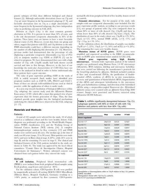
Table 1. miRNAs significantly <strong>de</strong>regulated between 13q- CLL<br />
subgroups (patients with 80% or more of cells with 13q<br />
<strong>de</strong>letion and patients with less than 80% 13q cells).<br />
miRNA Map q-value R fold<br />
Down-regulated<br />
Molecular Characterization of 13q- CLL<br />
hsa-mir-1-1* 20q13.33 0.0125 0.7027<br />
hsa-mir-7-1 9q21.32 0.0397 0.5453<br />
hsa-mir-15a 13q14.3 0.0329 0.4917<br />
hsa-mir-29a 7q32.3 0.0354 0.5101<br />
hsa-mir-34a* 1p36.23 0.0366 0.6874<br />
hsa-mir-106b* 7q22.1 0.0280 0.5190<br />
hsa-mir-181b 1q31.3 0.0256 0.6775<br />
hsa-mir-204 9q21.11 0.0294 0.5693<br />
hsa-mir-206 6p12.2 0.0476 0.7077<br />
hsa-mir-221* Xp11.3 0.0133 0.4622<br />
hsa-mir-223*<br />
Up-regulated<br />
Xq12 0.0017 0.1016<br />
hsa-mir-134 14q32.31 0.0095 1.8096<br />
hsa-mir-105-2 Xq28 0.0182 1.4040<br />
hsa-mir-155* 21q21.3 0.0046 3.7013<br />
hsa-mir-205 1q32.2 0.0161 1.3830<br />
Upregulation or downregulation refers to 13q-H relative to13q-L CLL patients.<br />
miRNA: microRNA.<br />
*<strong>de</strong>regulation shared with 17p/11q CLL patients.<br />
doi:10.1371/journal.pone.0048485.t001<br />
PLOS ONE | www.plosone.org 2 November 2012 | Volume 7 | Issue 11 | e48485


