Alberto Risueño Pérez - Gredos - Universidad de Salamanca
Alberto Risueño Pérez - Gredos - Universidad de Salamanca
Alberto Risueño Pérez - Gredos - Universidad de Salamanca

Create successful ePaper yourself
Turn your PDF publications into a flip-book with our unique Google optimized e-Paper software.
H patients. Moreover, our analysis showed an overexpression of<br />
genes promoting proliferation, such as LEF1, E2F5 and RRAS2.<br />
To ensure that the gene expression profiles accurately reflected the<br />
upregulation of BCR signaling pathway and the <strong>de</strong>regulation of<br />
apoptosis-related genes, representative genes that were differentially<br />
expressed in 13q-H patients were assessed by semiquantitative<br />
SYBRgreen PCR analysis. These inclu<strong>de</strong>d SYK,<br />
BLNK and PRKCB1 (BCR signaling pathway), BCL2 (apoptosis)<br />
and LEF1 and RRAS2 (proliferation). The semi-quantitative PCR<br />
results were in close agreement with the microarray data (Figure 1)<br />
confirming the overexpression of these genes in 13q-H CLLs<br />
compared with 13q-L. Western blot analysis should be ma<strong>de</strong> for a<br />
more concluding validation after mRNA screening. Unfortunately,<br />
due to the lack of material, this was not possible in this study.<br />
miRNA Deregulation in 13q-H CLL Patients<br />
The analysis of miRNA expression in 13q-H and 13q-L CLL<br />
patients revealed that fifteen miRNAs were <strong>de</strong>regulated in 13q-H<br />
CLL patients: hsa-miR-155 was the most highly upregulated<br />
miRNA (Rfold = 3.70), while hsa-miR-223 was the most significantly<br />
downregulated (Rfold = 0.10). Four of the <strong>de</strong>regulated<br />
miRNAs (miR-15a, miR-29a, miR-155 and miR- 223) were<br />
further assayed by quantitative RT-PCR for validation purposes in<br />
Molecular Characterization of 13q- CLL<br />
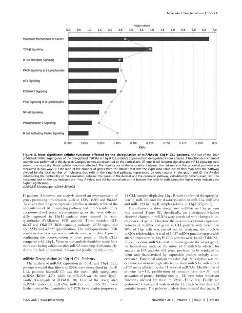
Figure 3. Most significant cellular functions affected by the <strong>de</strong>regulation of miRNAs in 13q-H CLL patients. 432 out of the 1027<br />
predicted mRNA target genes of the <strong>de</strong>regulated miRNAs in 13q-H CLL patients appeared also <strong>de</strong>regulated in our analysis. A functional enrichement<br />
analysis was performed in this dataset. Category names are presented on the vertical axis. Of note, B cell receptor signaling and NF-kB signaling were<br />
among the most significant cellular functions affected. The significance of the association between the dataset and the canonical pathway was<br />
measured in two ways: (1) the ratio of the number of genes from the dataset that met the expression value cut-off that map onto the pathway<br />
divi<strong>de</strong>d by the total number of molecules that exist in the canonical pathway, represented by grey squares in the graph and (2) the P-value<br />
<strong>de</strong>termining the probability of the association between the genes in the dataset and the canonical pathway, calculated by Fisher’s exact test. The<br />
horizontal axis on the top indicates the 2log (P value) and the horizontal axis at the bottom, the ratio. In both cases, the higher value indicates the<br />
higher significance.<br />
doi:10.1371/journal.pone.0048485.g003<br />
24 CLL samples displaying 13q-. Results confirmed the upregulation<br />
of miR-155 and the downregulation of miR-15a, miR-29a<br />
and miR- 223 in 13q-H samples relative to 13q-L (Figure 2).<br />
The influence of these <strong>de</strong>regulated miRNAs on 13q- patients<br />
was assessed (Figure S2). Specifically, we investigated whether<br />
observed changes in miRNAs were correlated with changes in the<br />
expression of genes. Therefore the post-transcriptional regulatory<br />
network of miRNA and genes in CLL patients with more than<br />
80% of 13q- cells was carried out by analyzing the miRNAmRNA<br />
relationships. A total of 1 027 mRNA putative targets with<br />
altered expression in 13q-H CLL patients were found (Table S4).<br />
In<strong>de</strong>ed, because miRNAs tend to downregulate the target genes,<br />
we focused our study on the subset of 11 miRNAs selected for<br />
analysis in IPA and the 432 genes predicted to be regulated by<br />
them and characterized by expression profiles stringly anticorrelated.<br />
Functional analysis revealed that transcription was the<br />
cell function most strongly affected by these miRNAs, with a total<br />
of 97 genes affected by the 11 selected miRNAs. Modification of<br />
proteins (n = 41), proliferation of immune cells (n = 34), and<br />
activation of protein binding sites (n = 32) were other important<br />
functions affected by these miRNAs (Table S5). Finally we<br />
performed a functional analysis of the 11 miRNAs and their 432<br />
putative targets. The pathway analysis <strong>de</strong>monstrated that, again, B<br />
PLOS ONE | www.plosone.org 7 November 2012 | Volume 7 | Issue 11 | e48485