Contribution à l'étude de virus de mollusques marins apparentés ...
Contribution à l'étude de virus de mollusques marins apparentés ...
Contribution à l'étude de virus de mollusques marins apparentés ...
Create successful ePaper yourself
Turn your PDF publications into a flip-book with our unique Google optimized e-Paper software.
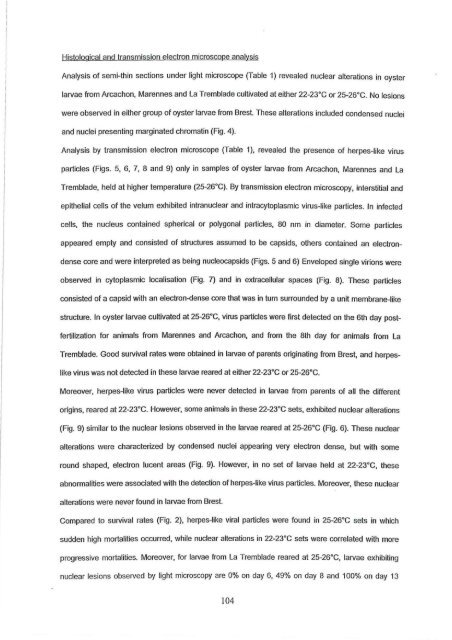
Histological and transmission electron microscope analysis<br />
Analysis of semi-thin sections un<strong>de</strong>r light microscope (Table 1) revealed nuclear alterations in oyster<br />
larvae trom Arcachon, Marennes and La Trembla<strong>de</strong> cultivated at either 22-23"C or 25-26"C. No les ions<br />
were observed in either group of oyster larvae trom Brest These alterations inclu<strong>de</strong>d con<strong>de</strong>nsed nuclei<br />
and nuclei presenting marginated chromatin (Fig. 4).<br />
Analysis by transmission electron microscope (Table 1), revealed the presence of herpes-like <strong>virus</strong><br />
particles (Figs. 5, 6, 7, 8 and 9) only in samples of oyster larvae from Arcachon, Marennes and La<br />
Trembla<strong>de</strong>, held at higher temperature (25-26"C). By transmission electron microscopy, interstitial and<br />
epithelial ce Us of the velum exhibited intranuclear and intracytoplasmic <strong>virus</strong>-like particles. In infected<br />
ceUs, the nucleus contained spherical or polygonal particles, 80 nm in diameter. Some particles<br />
appeared empty and consisted of structures assumed to be capsids, others contained an electron<br />
<strong>de</strong>nse core and were interpreted as being nucleocapsids (Figs. 5 and 6) Enveloped single virions were<br />
observed in cytoplasmic localisation (Fig. 7) and in extraceUular spaces (Fig. 8). These particles<br />
consisted of a capsid with an electron-<strong>de</strong>nse core that was in tum surroun<strong>de</strong>d by a unit membrane-like<br />
structure. In oyster larvae cultivated at 25-26"C, <strong>virus</strong> particles were first <strong>de</strong>tected on the 6th day post<br />
fertilization for animais from Marennes and Arcachon, and trom the 8th day for animais trom La<br />
Trembla<strong>de</strong>. Good survival rates were obtained in larvae of parents originating trom Brest, and herpes<br />
like <strong>virus</strong> was not <strong>de</strong>tected in these larvae reared at either 22-23"C or 25-26"C.<br />
Moreover, herpes-like <strong>virus</strong> particles were never <strong>de</strong>tected in larvae from parents of aU the different<br />
origins, reared at 22-23"C. However, some animais in these 22-23"C sets, exhibited nuclear alterations<br />
(Fig. 9) similar to the nuclear lesions observed in the larvae reared at 25-26"C (Fig. 6). These nuclear<br />
alterations were characterized by con<strong>de</strong>nsed nuclei appearing very electron <strong>de</strong>nse, but with some<br />
round shaped, electron lucent areas (Fig. 9). However, in no set of larvae he Id at 22-23"C, these<br />
abnorrnalities were associated with the <strong>de</strong>tection of herpes-like <strong>virus</strong> particles. Moreover, these nuclear<br />
alterations were never found in larvae from Brest.<br />
Compared to survival rates (Fig. 2), herpes-like viral particles were found in 25-26"C sets in which<br />
sud<strong>de</strong>n high mortalities occurred, while nuclear alterations in 22-23"C sets were correlated with more<br />
progressive mortalities. Moreover, for larvae trom La Trembla<strong>de</strong> reared at 25-26"C, larvae exhibiting<br />
nuclear les ions observed by light microscopy are 0% on day 6, 49% on day 8 and 100% on day 13<br />
104


