Contribution à l'étude de virus de mollusques marins apparentés ...
Contribution à l'étude de virus de mollusques marins apparentés ...
Contribution à l'étude de virus de mollusques marins apparentés ...

Create successful ePaper yourself
Turn your PDF publications into a flip-book with our unique Google optimized e-Paper software.
PUBLICATION 7<br />
Journal Q/Tissue CI/lIUre Methods 16: 67-72. 1994.<br />
© 1994 Kl/IIH'r Aca<strong>de</strong>mie Publishers. Primed i" the Netlurlands.<br />
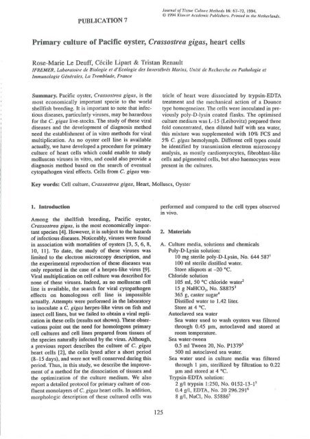
Primary culture of Pacific oyster, Crassostrea gigas, heart cells<br />
Rose-Marie Le Deuff, Cécile Lipart & Tristan Renault<br />
lFREMER, Laboratoire <strong>de</strong> Biologie et d'Ecologie <strong>de</strong>s Invertébrés Marins, Unité <strong>de</strong> Recherche en Pathologie et<br />
Immunologie Générales. La Trembla<strong>de</strong>, France<br />
Summary. Pacific oyster, Crassostrea gigas, is the<br />
most economicaIly important specie to the world<br />
sheIlfish breeding. It is important to note that infec·<br />
tious diseases, particularly <strong>virus</strong>es, may be hazardous<br />
for the C. gigas live-stocks. The study of these viral<br />
di seases and the <strong>de</strong>velopment of diagnosis method<br />
need the establishment of in vitro methods for viral<br />
multiplication. As no oyster œIl line is available<br />
actually, we have <strong>de</strong>veloped a procedure for primary<br />
culture of heart ceIls which could enable to study<br />
molluscan <strong>virus</strong>es in vitro, and could also provi<strong>de</strong> a<br />
diagnosis method based on the search of eventual<br />
cytopathogen viral effects. Cells from C. gigas ven-<br />
Key words: Cell culture, Crassostrea gigas, Heart, MoIluscs, Oyster<br />
1. Introduction<br />
Among the shellfish breeding, Pacific oyster,<br />
Crassostrea gigas, is the most economicaIly important<br />
species (4), However, it is subject to the hazards<br />
of infectious diseases. Noticeably, <strong>virus</strong>es were found<br />
in association with mortalities of oysters [3, 5, 6, 8,<br />
10, Il). To date, the study of these <strong>virus</strong>es was<br />
limited to the electron microscopy <strong>de</strong>scription, and<br />
the experimental reproduction of these diseases was<br />
only reported in the case of a herpes-like <strong>virus</strong> (9).<br />
Viral multiplication on cell culture was <strong>de</strong>scribed for<br />
none of these <strong>virus</strong>es. In<strong>de</strong>ed, as no molluscan œil<br />
line is available, the search for viral cytopathogen<br />
effects on homologous cell line is impossible<br />
actually. Attempts were perfonmed in the laboratory<br />
to inoculate a C. gigas herpes-Iike <strong>virus</strong> on fish and<br />
insect œlllines, but we failed to obtain a viral replication<br />
in these cells (results not shown). These observations<br />
point out the need for homologous primary<br />
ceIl cultures and cell Iines prepared from tissues of<br />
the species naturaIly infected by the <strong>virus</strong>. Although,<br />
a previous report <strong>de</strong>scribes the culture of C. gigas<br />
heart cells (2), the cellS Iysed after a short period<br />
(8- 15 days), and were not weil conserved during this<br />
period. Thus, in this study, we <strong>de</strong>scribe the improvement<br />
of a method for the dissociation of tissues and<br />
the optimization of the culture medium. We also<br />
report a <strong>de</strong>tailed protocol for primary culture of confluent<br />
monolayers of C. gigas heart cells. In addition,<br />
morphologic <strong>de</strong>scription of Ihese cultured ceIls was<br />
125<br />
tricle of heart were dissociated by trypsin-EDTA<br />
treatment and the mechanical action of a Dounce<br />
type homogeneizer. The ceIls were inoculated in previously<br />
poly-D-Iysin coated flasks. The optimised<br />
culture medium was L-15 (Leibovitz) prepared three<br />
fold concentrated. then diluted half with sea water,<br />
this mixture was supplemented with 10% FCS and<br />
5% C. gigas hemolymph. Different cell types could<br />
be i<strong>de</strong>ntified by transmission electron microscopy<br />
analysis, as mostly cardiomyocytes, fibroblast-like<br />
ceIls and pigmented cells, but also haemocytes were<br />
present in the cultures.<br />
performed and compared to the œil types observed<br />
in vivo.<br />
2. Materials<br />
A. Culture media, solutions and chemicals<br />
Poly-D-Lysin solution:<br />
10 mg sterile poly-D-Lysin, No. 644 587'<br />
100 ml sterile distilled water.<br />
Store aliquots al -20 oC,<br />
Chlori<strong>de</strong> solution<br />
105 ml, 50 oC chlori<strong>de</strong> water'<br />
15 g NaHCO" No. S8875'<br />
365 g. caster sugar'<br />
DistiIled water to 1.42 liter.<br />
Store at 4 oC.<br />
Autoc1aved sea water<br />
Sea water used to wash oysters was filtered<br />
through 0.45 !lm, autoc1aved and stored at<br />
room temperature.<br />
Sea water-tween<br />
0.5 ml Tween 20, No. PI379'<br />
500 ml autoc1aved sea water.<br />
Sea water used in culture media was filtered<br />
through 1 !lm, sterilized by filtration to 0.22<br />
!lm and stored at 4 oC,<br />
Trypsin-EDTA solution:<br />
2 gll trypsin 1:250, No. 0152-13-1'<br />
0.4 gll, EDTA, No. 20 296.291 6<br />
8 gll. NaCI, No. S5886'