

QF0159 Marketing Release Record
QF0159 Marketing Release Record
QF0159 Marketing Release Record

Create successful ePaper yourself
Turn your PDF publications into a flip-book with our unique Google optimized e-Paper software.
Primary Antibodies<br />
Novocastra Tissue Inhibitor of Matrix<br />
Metalloproteinase Antibodies<br />
Clone 6F6a<br />
1 mL lyophilized Tissue Inhibitor of Matrix<br />
Metalloproteinase 1 NCL-TIMP1-485 P (HIER) W<br />
Clone 46E5<br />
1 mL lyophilized Tissue Inhibitor of Matrix<br />
Metalloproteinase 2 NCL-TIMP2-487 P (HIER) W<br />
Clone 18D12b<br />
1 mL lyophilized Tissue Inhibitor of Matrix<br />
Metalloproteinase 3 NCL-TIMP3 FP RUO*<br />
The tissue inhibitors of metalloproteinases (TIMPs) are natural inhibitors of<br />
matrix metalloproteinases (MMPs), a group of zinc-binding endopeptides<br />
involved in degradation of the extracellular matrix (ECM). The remodelling of<br />
the ECM in a controlled fashion is essential during normal development and<br />
is a feature of physiological remodelling such as in wound healing. Tumor<br />
cell invasion and metastasis closely correlate with the activities of two<br />
MMPs, MMP2 and MMP9, both of which degrade type IV collagen in<br />
basement membranes. TIMPs constitute a family of at least four types of<br />
protein of which two of these are expressed in a wide variety of cell types.<br />
Although TIMP2 inhibits all types of activated MMPs to varying degrees, it is<br />
known to preferentially inhibit MMP2. TIMP2 also binds to proMMP9 and<br />
proMMP2 to form stable complexes in which activation of the proenzymes<br />
occur. Studies have revealed that TIMP2 can inhibit the invasive potential of<br />
tumor cells in vitro and their metastatic phenotype in vivo. TIMP3 is secreted<br />
into the ECM and complexes with MMP1, 3, 7, 9, 13, 14 and 15 deactivating<br />
them irreversibly. TIMP3 mRNA is highly expressed in placenta but is also<br />
reported to be found in the heart, kidney, lung, pancreas, uterus and skeletal<br />
muscle with low levels in the brain. In early placenta, TIMP2 and TIMP3<br />
mRNAs are reported to be found in cells of cytotrophoblastic columns and<br />
decidual membrane. In endometrium, TIMP3 is reported to be expressed in<br />
luminal epithelium, glands, stroma, endothelial cells and vascular smooth<br />
muscle cells. In adult rat cerebellum, TIMP3 is reported to be expressed in<br />
Purkinje cell somata and processes. TIMP3 is expressed by fibroblast-like<br />
cells in ulcerated intestinal wall and in surrounding vessels and sweat<br />
glands during wound healing in skin. In breast carcinoma, TIMP3 mRNA<br />
expression is described in fibroblastic cells within the tumor stroma<br />
adjacent to cancer cells.<br />
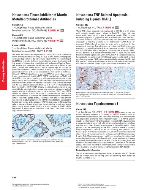
Human small intestine: immunohistochemical staining for tissue inhibitor of matrix<br />
metalloproteinase 1 using NCL-TIMP1-485. Note cytoplasmic staining of epithelial<br />
cells. Paraffin section.<br />
/ 106<br />
For detailed information on all products please visit our website:<br />
www.leica-microsystems.com<br />
RUO*<br />
RUO*<br />
Novocastra TNF-Related Apoptosis-<br />
Inducing Ligand (TRAIL)<br />
Clone 27B12<br />
1 mL lyophilized NCL-TRAIL P (HIER) W<br />
TRAIL (TNF-related apoptosis-inducing ligand), or APO-2L, is a 281 amino<br />
acid cytotoxic protein closely related to Fas/APO-1 ligand with the<br />
characteristic structure of a type II membrane protein. TRAIL induces<br />
extensive apoptosis in lymphoid as well as nonlymphoid tumor cell lines.<br />
Two TRAIL membrane receptors, DR4 and DR5, have been identified which<br />
are capable of mediating apoptosis and are distinct from Fas/APO-1 and TNF<br />
receptors. TRAIL-induced apoptosis in target cells is mediated by the<br />
activation of caspases. Normal tissues are resistant to TRAIL as they are<br />
reported to express high levels of decoy membrane receptors, DcR1/TRID<br />
and DcR2/TRUNDD which antagonize TRAIL-induced apoptosis. TRAIL<br />
induces apoptosis in a number of different tumor cell types because most<br />
transformed cells express little DcR1. TRAIL mRNA is expressed in spleen,<br />
lung, prostate, ovary and bowel with little expression in testis, heart, skeletal<br />
muscle and pancreas. TRAIL protein is reported to be expressed on CD4 and<br />
CD8 positive T lymphocytes following activation and is also predominant in<br />
first trimester placental syncytiotrophoblasts as well as Hofbauer cells.<br />
Human prostatic carcinoma: immunohistochemical staining for TNF-related apoptosisinducing<br />
ligand using NCL-TRAIL. Note cytoplasmic staining of a proportion of malignant<br />
cells. Paraffin section.<br />
Novocastra Topoisomerase I<br />
Clone 1D6<br />
1 mL lyophilized NCL-TOPO I F P (HIER) Topoisomerases are<br />
nuclear enzymes involved in a variety of cellular activities such as<br />
chromosomal condensation, DNA replication, transcrip-tion, recombination<br />
and segregation at mitosis. Human topoisomerase I is a 100 kD protein<br />
capable of relaxing positively and negatively supercoiled DNA by performing<br />
a transient single-stranded nick which is then re-ligated at the end of the<br />
reaction. It has been shown that the enzyme is located in regions of the<br />
genome that are undergoing active RNA synthesis where it probably<br />
reduces superhelical stresses in the DNA enabling RNA polymerase to<br />
function properly. In normal eukaryotic cells, DNA topoisomerase I does not<br />
show relevant fluctuations across the cell cycle, unlike DNA topoisomerase<br />
II alpha. Both DNA topoisomerases I and II have been found to be targets of<br />
autoantibodies in the sera of individuals with certain autoimmune diseases<br />
eg systemic lupus erythematosus and also of some anti-tumor drugs and<br />
antibiotics. Elevated levels of DNA topoisomerase I, detected by 32 IVD<br />
P transfer<br />
assays, have been reported in colorectal tumors compared with normal<br />
colon mucosa as a result of increased transcription or mRNA stability.<br />
Product Specific Information<br />
The use of phosphate-containing wash buffers or diluents with NCL-TOPO I<br />
has an adverse effect on staining. Only Tris-containing wash buffers or<br />
diluents should be used.<br />
Products in this catalog are subject to regulatory approval.<br />
Please consult your Leica Microsystems representative for availability in your region.<br />
* For Research Use Only. Not for use in diagnostic procedures.<br />
RUO*


