

QF0159 Marketing Release Record
QF0159 Marketing Release Record
QF0159 Marketing Release Record

You also want an ePaper? Increase the reach of your titles
YUMPU automatically turns print PDFs into web optimized ePapers that Google loves.
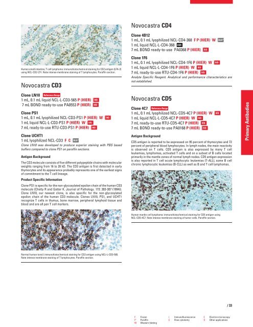
Human small intestine, T cell lymphoma: immunohistochemical staining for CD2 antigen (LFA-2)<br />
using NCL-CD2-271. Note intense membrane staining of T lymphocytes. Paraffin section.<br />
Novocastra CD3<br />
Clone LN10 Reference Range<br />
1 mL, 0.1 mL liquid NCL-L-CD3-565 P (HIER)<br />
7 mL BOND ready-to-use PA0553 P (HIER)<br />
Clone PS1<br />
1 mL, 0.1 mL lyophilized NCL-CD3-PS1 P (HIER) W<br />
1 mL liquid NCL-L-CD3-PS1 P (HIER) W IVD<br />
7 mL ready-to-use RTU-CD3-PS1 P (HIER) IVD<br />
Clone UCHT1<br />
1 mL lyophilized NCL-CD3 FC RUO*<br />
Clone LN10 was developed to produce superior staining with PBS based<br />
buffers compared to clone PS1 on paraffin sections.<br />
Antigen Background<br />
The CD3 molecule consists of five different polypeptide chains with molecular<br />
weights ranging from 16 to 28 kD. The CD3 antigen is first detected in early<br />
thymocytes and its appearance probably represents one of the earliest signs<br />
of commitment to the T cell lineage.<br />
Product Specific Information<br />
Clone PS1 is specific for the non-glycosylated epsilon chain of the human CD3<br />
molecule (Chetty R and Gatter K. Journal of Pathology. 173: 303-307 (1994)).<br />
Clone LN10, our newest clone, is also specific for the non-glycosylated<br />
epsilon chain of the human CD3 molecule. Clones LN10, PS1, and UCHT1<br />
recognize T cells in thymus, bone marrow, peripheral lymphoid tissue and<br />
blood and are all pan T cell markers.<br />
Normal human tonsil: immunohistochemical staining for CD3 antigen using NCL-L-CD3-565.<br />
Note intense membrane staining of T lymphocytes. Paraffin section.<br />
IVD<br />
IVD<br />
IVD<br />
Novocastra CD4<br />
Clone 4B12<br />
1 mL, 0.1 mL lyophilized NCL-CD4-368 F P (HIER) W<br />
1 mL liquid NCL-L-CD4-368 ASR<br />
7 mL BOND ready-to-use PA0368 P (HIER) IVD<br />
Clone 1F6<br />
1 mL, 0.1 mL lyophilized NCL-CD4-1F6 P (HIER) W IVD<br />
1 mL liquid NCL-L-CD4-1F6 P (HIER) W IVD<br />
7 mL ready-to-use RTU-CD4-1F6 P (HIER) IVD<br />
Analyte Specific Reagent. Analytical and performance characteristics are<br />
not established.<br />
Novocastra CD5<br />
Clone 4C7 Reference Range<br />
1 mL, 0.1 mL lyophilized NCL-CD5-4C7 P (HIER) W<br />
1 mL liquid NCL-L-CD5-4C7 P (HIER) W IVD<br />
7 mL ready-to-use RTU-CD5-4C7 P (HIER) IVD<br />
7 mL BOND ready-to-use PA0168 P (HIER) IVD<br />
Antigen Background<br />
CD5 antigen is reported to be expressed on 95 percent of thymocytes and 72<br />
percent of peripheral blood lymphocytes. In lymph nodes, the main reactivity<br />
is observed on T cells. CD5 antigen is also expressed by many T cell<br />
leukemias, lymphomas, activated T cells and on a subset of B cells located<br />
primarily in the mantle zones of normal lymph nodes. CD5 antigen expression<br />
is also reported in T cell acute lymphocytic leukemias (T-ALL), some B cell<br />
chronic lymphocytic leukemias (B-CLL) as well as B and T cell lymphomas.<br />
Human mantle cell lymphoma: immunohistochemical staining for CD5 antigen using<br />
NCL-CD5-4C7. Note intense membrane staining of tumor cells. Paraffin section.<br />
RUO*<br />
F Frozen I Immunofluorescence E Electron microscopy<br />
P Paraffin C Flow cytometry O Other applications<br />
W Western blotting<br />
IVD<br />
/33<br />
Primary Antibodies


