

QF0159 Marketing Release Record
QF0159 Marketing Release Record
QF0159 Marketing Release Record

You also want an ePaper? Increase the reach of your titles
YUMPU automatically turns print PDFs into web optimized ePapers that Google loves.
Novocastra CD69<br />
Clone CH11<br />
1 mL lyophilized NCL-CD69 P (HIER)<br />
The CD69 molecule is a type II membrane glycoprotein expressed as a<br />
disulfide-linked homodimer. The human and mouse genes for CD69 are<br />
encoded within the NK gene complex on chromosomes 12 and 6,<br />
respectively. CD69 protein is expressed mainly on activated T and<br />
B lymphocytes.<br />
Human tonsil: immunohistochemical staining for CD69 antigen using NCL-CD69. Note<br />
membrane staining of activated lymphocytes, NK cells and neutrophils. Paraffin section.<br />
Novocastra CD71<br />
Clone 10F11<br />
1 mL lyophilized NCL-CD71-309 P (HIER)<br />
The CD71 molecule is a type II membrane glycoprotein with a molecular<br />
weight of approximately 180 kD. It is known as the transferrin receptor and is<br />
composed of two disulfide bonded 90 kD subunits. The CD71 molecule plays<br />
a critical role in cell proliferation by controlling the supply of iron, an<br />
essential component for many metabolic pathways, through the binding and<br />
endocytosis of transferrin, the major iron-carrying protein. CD71 protein is<br />
reported to be expressed on activated B and T cells, macrophages,<br />
proliferating cells and metabolically active cells eg neurons.<br />
Novocastra CD74<br />
Clone LN-2<br />
1 mLlyophilized NCL-LN2 F P (HIER)<br />
Novocastra CD75<br />
Clone 11E3<br />
1 mL lyophilized NCL-LN1 F P (HIER)<br />
RUO*<br />
RUO*<br />
RUO*<br />
Human centroblastic lymphoma: immunohistochemical staining for CD75 antigen using<br />
NCL-LN1. Note intense membrane staining of B lymphocytes. Paraffin section.<br />
IVD<br />
Novocastra CD79a<br />
Clone 11E3 Reference Range<br />
1 mL, 0.1 mL lyophilized NCL-CD79a-225 F P (HIER)<br />
1 mL liquid NCL-L-CD79a-225 F P (HIER) C IVD<br />
7 mL BOND ready-to-use PA0192 P (HIER) IVD<br />
Clone 11D10<br />
1 mL, 0.1 mL lyophilized NCL-CD79a-192 F P (HIER) C RUO*<br />
1 mL liquid NCL-L-CD79a-192 F P (HIER) RUO*<br />
7 mL ready-to-use RTU-CD79a-192 F P (HIER) RUO*<br />
Clone 11E3 was developed to produce superior staining on paraffin sections.<br />
Antigen Background<br />
The CD79 complex is a disulfide-linked heterodimer which is non-covalently<br />
associated with membrane-bound immunoglobulins on B cells. This complex<br />
of polypeptides and immunoglobulin constitute the B cell antigen receptor.<br />
The two components of this complex are designated CD79a and CD79b. The<br />
CD79a antigen is reported to first appear at the pre-B cell stage, early in<br />
maturation, and persist until the plasma cell stage where it is found as an<br />
intracellular component. The CD79a antigen is reported to be expressed in<br />
the majority of acute leukemias of precursor B cell type, B cell lines, B cell<br />
lymphomas and in some myelomas. It is not present in myeloid or T cell lines.<br />
Human large cell lymphoma: immunohistochemical staining for CD79a antigen using<br />
NCL-CD79a-225. Note membrane staining of tumor cells. Paraffin section.<br />
Novocastra CD79b<br />
Clone JS01<br />
1 mL, 0.1 mL liquid NCL-L-CD79b P (HIER)<br />
RUO*<br />
CD79b, also known as B29 and Ig-� is thought to function in the cellular<br />
activation and signalling that occurs when surface immunoglobulin (Ig) on B<br />
cells binds antigen or becomes cross-linked by anti-Ig antibody. This<br />
function occurs with the formation of a membrane signalling complex that is<br />
associated with Ig at the surface of B cells. CD79b, together with CD79a,<br />
forms the B cell antigen receptor (mlg) complex. CD79b expression is<br />
reported to be found in 80 to 90 percent of mature B cell neoplasms, with the<br />
exception of chronic lymphocytic leukemias.<br />
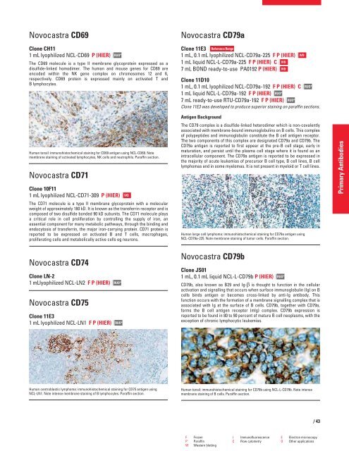
Human tonsil: immunohistochemical staining for CD79b using NCL-L-CD79b. Note intense<br />
membrane staining of B cells. Paraffin section.<br />
F Frozen I Immunofluorescence E Electron microscopy<br />
P Paraffin C Flow cytometry O Other applications<br />
W Western blotting<br />
IVD<br />
/43<br />
Primary Antibodies


