

QF0159 Marketing Release Record
QF0159 Marketing Release Record
QF0159 Marketing Release Record

Create successful ePaper yourself
Turn your PDF publications into a flip-book with our unique Google optimized e-Paper software.
Human Colonic Carcinoma: immunohistochemical staining for PMS2 using NCL-L-PMS2.<br />
Paraffin section.<br />
Novocastra Motility-Related Protein-1<br />
(CD9)<br />
Clone 72F6<br />
1 mL lyophilized NCL-CD9 F P (HIER)<br />
See also CD9 (Motility-Related Protein-1) on page 34.<br />
Novocastra Muc Glycoprotein<br />
Antibodies<br />
Clone Ma552<br />
1 mL lyophilized muc-1 core glycoprotein<br />
NCL-MUC-1-CORE F P (HIER) RUO*<br />
Clone Ma695<br />
1 mL lyophilized muc-1 glycoprotein<br />
NCL-MUC-1 F P (HIER) IVD<br />
Clone Ccp58<br />
1 mL, 0.1 mL lyophilized muc-2 glycoprotein<br />
NCL-MUC-2 F P (HIER) IVD<br />
Clone CLH2<br />
1 mL, 0.1 mL lyophilized muc-5AC glycoprotein<br />
NCL-MUC-5AC P (HIER) IVD<br />
Clone CLH5<br />
1 mL, 0.1 mL lyophilized muc-6 glycoprotein<br />
NCL-MUC-6 P (HIER) IVD<br />
Antigen Background<br />
RUO*<br />
Mucins are heavily glycosylated proteins which constitute the major<br />
components of mucus covering the surface of epithelial tissues. Nine distinct<br />
epithelial mucin genes (Muc-1, 2, 3, 4, 5AC, 5B, 6, 7 and 8) have been<br />
identified. Various immunohistochemical and in situ hybridization studies<br />
have reported that these mucins are differentially expressed in epithelia with<br />
cell-type specificity. The normal gastric mucosa shows cell-type specific<br />
expression of Muc-1, Muc-5AC and Muc-6 glycoproteins. Muc-1 and Muc-<br />
5AC are found in superficial epithelium and Muc-6 glycoprotein in the deep<br />
glands. Muc-1 and Muc-5AC glycoproteins are reported to be expressed in<br />
many epithelia but Muc-6 glycoprotein is mainly expressed in gastric<br />
mucosa. In addition, Muc-2 glycoprotein is not expressed in normal gastric<br />
mucosa. In gastric cancer, alterations in mucin polypeptide expression have<br />
been reported, including the loss of expression of Muc-5AC glycoprotein,<br />
increased mucin heterogeneity, glycosylation changes and the expression of<br />
simple mucin-type carbohydrates.<br />
Normal human stomach: immunohistochemical staining for Muc-6 glycoprotein using NCL-<br />
MUC-6. Note cytoplasmic staining of mucus secreting cells of the deep glands. Paraffin<br />
section.<br />
Novocastra Multiple Myeloma<br />
Oncogene 1 (MUM-1)<br />
Clone EAU32<br />
1 mL, 0.1 mL liquid NCL-L-MUM1 P (HIER)<br />
7 mL BOND ready-to-use PA0129 P (HIER)<br />
Antigen Background<br />
The MUM-1 (multiple myeloma oncogene 1) gene was originally identified<br />
because of it’s involvement in the t(6:14) translocation observed in multiple<br />
myeloma, which causes the juxtaposition of the MUM-1 gene to the Ig heavy<br />
chain locus. MUM-1 is expressed in late plasma cell directed stages of B cell<br />
differentiation and in activated T cells, suggesting that MUM-1 may serve as<br />
a marker for lympho-hemopoetic neoplasms derived from these cells. The<br />
morphologic spectrum of MUM-1 expressing cells has been found to range<br />
from that of a centrocyte to that of a plasmablast/plasma cell. Consequently<br />
the histogenic value of MUM-1 may be to provide a marker to aid in the<br />
identification of the transition from BCL-6 positive (germinal center B cells) to<br />
CD138 positive (immunoblasts and plasma cells). MUM-1 expression occurs<br />
in a wide range of lymphoid neoplasms including a proportion of diffuse B cell<br />
lymphomas but not myeloid or extra-hemopoietic neoplasms. MUM-1 is<br />
consistently expressed in myeloma cells, Reed Sternberg cells in classic<br />
Hodgkin Disease, and activated and neoplastic T cells.<br />
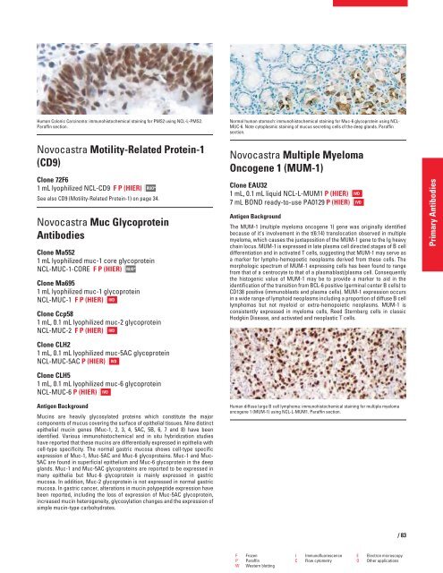
Human diffuse large B cell lymphoma: immunohistochemical staining for multiple myeloma<br />
oncogene 1 (MUM-1) using NCL-L-MUM1. Paraffin section.<br />
F Frozen I Immunofluorescence E Electron microscopy<br />
P Paraffin C Flow cytometry O Other applications<br />
W Western blotting<br />
IVD<br />
IVD<br />
/83<br />
Primary Antibodies


