

QF0159 Marketing Release Record
QF0159 Marketing Release Record
QF0159 Marketing Release Record

Create successful ePaper yourself
Turn your PDF publications into a flip-book with our unique Google optimized e-Paper software.
Novocastra Factor XIIIa (Blood<br />
Coagulation Factor XIIIa)<br />
Clone E980.1<br />
1 mL lyophilized NCL-FXIIIa P (HIER) IVD<br />
7 mL BOND ready-to-use PA0449 P (HIER)<br />
Antigen Background<br />
Factor XIIIa also known as fibrinoligase and fibrin-stabilizing factor, is the last<br />
enzyme generated in the blood coagulation cascade. It is a Ca 2+ - dependent<br />
transglutaminase or transamidating enzyme which forms intermolecular<br />
gamma-glutamyl-epsilon-lysine crosslinks between fibrin molecules<br />
resulting in the mechanical stabilization of the fibrin clot and its resistance to<br />
proteolysis. Factor XIIIa may also function to stabilize cell surface molecules<br />
and membranes. These Ca 2+ -dependent trans-glutaminases with thiol active<br />
centers are widespread in animal tissues and have been associated with cell<br />
proliferation, embryonic development and growth through the proliferation of<br />
mammary stroma and epithelial elements. Normal mammary stroma, like<br />
most collagenous connective tissue contains resident populations of CD34<br />
positive dendritic interstitial cells and scattered factor XIIIa positive<br />
collagen-associated dendrophages. Factor XIIIa has been examined to<br />
determine its expression in normal and inflamed skin. Factor XIIIa positive<br />
cells in human skin represent a specific population of bone marrow dermal<br />
dendritic cells, distinct from Langerhans cells which share some features<br />
common to mononuclear phagocytes. In benign skin conditions such as<br />
inflammatory dermatoses eg atopic eczema and psoriasis, an increased<br />
number of factor XIIIa positive cells in the upper dermis, closely associated<br />
with lymphocytes, has been described.<br />
Novocastra Fas (CD95)<br />
Clone GM30<br />
1 mL lyophilized NCL-FAS-310 F P (HIER)<br />
Fas is a 48 kD transmembrane glycoprotein. It is a member of the nerve<br />
growth factor receptor/tumor necrosis factor superfamily. This cell surface<br />
molecule mediates receptor-triggered apoptosis (programmed cell death).<br />
During embryonic and postembryonic development, many cells die by<br />
means of apoptosis. This plays a major role in determining morphological<br />
and functional maturity in a variety of systems, including the formation of the<br />
neural network and clonal deletion of autoreactive T cells. Apoptosis is<br />
accompanied by condensation of the cytoplasm, loss of plasma membrane<br />
microvilli and extensive degradation of chromosomal DNA into oligomers of<br />
about 180 base pairs. The Fas antigen is reported to be expressed on the<br />
surface of various cell types, including activated T and B lymphocytes and<br />
T lymphoblastoid cell lines.<br />
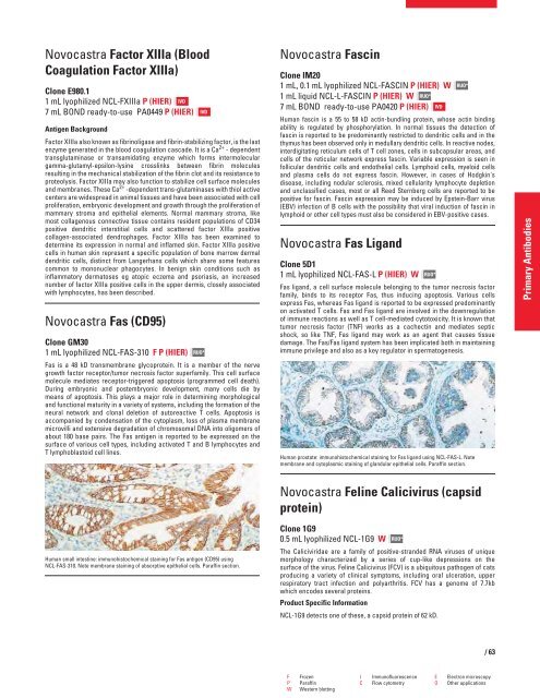
Human small intestine: immunohistochemical staining for Fas antigen (CD95) using<br />
NCL-FAS-310. Note membrane staining of absorptive epithelial cells. Paraffin section.<br />
IVD<br />
RUO*<br />
Novocastra Fascin<br />
Clone IM20<br />
1 mL, 0.1 mL lyophilized NCL-FASCIN P (HIER) W<br />
1 mL liquid NCL-L-FASCIN P (HIER) W RUO*<br />
7 mL BOND ready-to-use PA0420 P (HIER) IVD<br />
Human fascin is a 55 to 58 kD actin-bundling protein, whose actin binding<br />
ability is regulated by phosphorylation. In normal tissues the detection of<br />
fascin is reported to be predominantly restricted to dendritic cells and in the<br />
thymus has been observed only in medullary dendritic cells. In reactive nodes,<br />
interdigitating reticulum cells of T cell zones, cells in subcapsular areas, and<br />
cells of the reticular network express fascin. Variable expression is seen in<br />
follicular dendritic cells and endothelial cells. Lymphoid cells, myeloid cells<br />
and plasma cells do not express fascin. However, in cases of Hodgkin's<br />
disease, including nodular sclerosis, mixed cellularity lymphocyte depletion<br />
and unclassified cases, most or all Reed Sternberg cells are reported to be<br />
positive for fascin. Fascin expression may be induced by Epstein-Barr virus<br />
(EBV) infection of B cells with the possibility that viral induction of fascin in<br />
lymphoid or other cell types must also be considered in EBV-positive cases.<br />
Novocastra Fas Ligand<br />
Clone 5D1<br />
1 mL lyophilized NCL-FAS-L P (HIER) W<br />
Fas ligand, a cell surface molecule belonging to the tumor necrosis factor<br />
family, binds to its receptor Fas, thus inducing apoptosis. Various cells<br />
express Fas, whereas Fas ligand is reported to be expressed predominantly<br />
on activated T cells. Fas and Fas ligand are involved in the downregulation<br />
of immune reactions as well as T cell-mediated cytotoxicity. It is known that<br />
tumor necrosis factor (TNF) works as a cachectin and mediates septic<br />
shock, so like TNF, Fas ligand may work as an agent that causes tissue<br />
damage. The Fas/Fas ligand system has been implicated both in maintaining<br />
immune privilege and also as a key regulator in spermatogenesis.<br />
Human prostate: immunohistochemical staining for Fas ligand using NCL-FAS-L. Note<br />
membrane and cytoplasmic staining of glandular epithelial cells. Paraffin section.<br />
Novocastra Feline Calicivirus (capsid<br />
protein)<br />
Clone 1G9<br />
0.5 mL lyophilized NCL-1G9 W<br />
RUO*<br />
RUO*<br />
The Caliciviridae are a family of positive-stranded RNA viruses of unique<br />
morphology characterized by a series of cup-like depressions on the<br />
surface of the virus. Feline Calicivirus (FCV) is a ubiquitous pathogen of cats<br />
producing a variety of clinical symptoms, including oral ulceration, upper<br />
respiratory tract infection and polyarthritis. FCV has a genome of 7.7kb<br />
which encodes several proteins.<br />
Product Specific Information<br />
NCL-1G9 detects one of these, a capsid protein of 62 kD.<br />
RUO*<br />
F Frozen I Immunofluorescence E Electron microscopy<br />
P Paraffin C Flow cytometry O Other applications<br />
W Western blotting<br />
/63<br />
Primary Antibodies


