

QF0159 Marketing Release Record
QF0159 Marketing Release Record
QF0159 Marketing Release Record

Create successful ePaper yourself
Turn your PDF publications into a flip-book with our unique Google optimized e-Paper software.
Novocastra CD163<br />
Clone 10D6<br />
1 mL lyophilized NCL-CD163 P (HIER)<br />
Antigen Background<br />
The CD163 molecule is a type I membrane protein also known as M130<br />
antigen, Ber-Mac3, Ki-M8 or SM4. CD163 protein is restricted in its<br />
expression to the monocytic/macrophage lineage. It is reported to be present<br />
on all circulating monocytes and most tissue macrophages except those<br />
found in the mantle zone and germinal centers of lymphoid follicles,<br />
interdigitating reticulum cells and Langerhans cells. In addition, multinucleated<br />
cells within inflammatory lesions are reported not to express<br />
CD163 protein. The protein is upregulated by glucocorticoids and<br />
downregulated by the immunosuppressant cyclosporin A and by phorbol<br />
esters, while lipopolysaccharide, an inflammatory mediator, has no influence<br />
on expression. It has been proposed that a specific release mechanism of<br />
soluble CD163 antigen by human monocytes may play an important role in<br />
modulating inflammatory processes.<br />
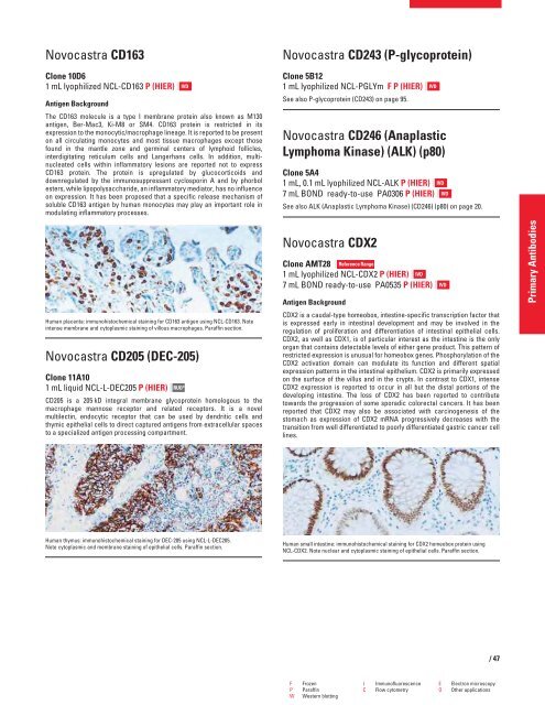
Human placenta: immunohistochemical staining for CD163 antigen using NCL-CD163. Note<br />
intense membrane and cytoplasmic staining of villous macrophages. Paraffin section.<br />
Novocastra CD205 (DEC-205)<br />
Clone 11A10<br />
1 mL liquid NCL-L-DEC205 P (HIER)<br />
RUO*<br />
CD205 is a 205 kD integral membrane glycoprotein homologous to the<br />
macrophage mannose receptor and related receptors. It is a novel<br />
multilectin, endocytic receptor that can be used by dendritic cells and<br />
thymic epithelial cells to direct captured antigens from extracellular spaces<br />
to a specialized antigen processing compartment.<br />
Human thymus: immunohistochemical staining for DEC-205 using NCL-L-DEC205.<br />
Note cytoplasmic and membrane staining of epithelial cells. Paraffin section.<br />
IVD<br />
Novocastra CD243 (P-glycoprotein)<br />
Clone 5B12<br />
1 mL lyophilized NCL-PGLYm F P (HIER)<br />
See also P-glycoprotein (CD243) on page 95.<br />
Novocastra CD246 (Anaplastic<br />
Lymphoma Kinase) (ALK) (p80)<br />
Clone 5A4<br />
1 mL, 0.1 mL lyophilized NCL-ALK P (HIER)<br />
7 mL BOND ready-to-use PA0306 P (HIER)<br />
See also ALK (Anaplastic Lymphoma Kinase) (CD246) (p80) on page 20.<br />
Novocastra CDX2<br />
Clone AMT28 Reference Range<br />
1 mL lyophilized NCL-CDX2 P (HIER) IVD<br />
7 mL BOND ready-to-use PA0535 P (HIER)<br />
Antigen Background<br />
CDX2 is a caudal-type homeobox, intestine-specific transcription factor that<br />
is expressed early in intestinal development and may be involved in the<br />
regulation of proliferation and differentiation of intestinal epithelial cells.<br />
CDX2, as well as CDX1, is of particular interest as the intestine is the only<br />
organ that contains detectable levels of either gene product. This pattern of<br />
restricted expression is unusual for homeobox genes. Phosphorylation of the<br />
CDX2 activation domain can modulate its function and different spatial<br />
expression patterns in the intestinal epithelium. CDX2 is primarily expressed<br />
on the surface of the villus and in the crypts. In contrast to CDX1, intense<br />
CDX2 expression is reported to occur in all but the distal portions of the<br />
developing intestine. The loss of CDX2 has been reported to contribute<br />
towards the progression of some sporadic colorectal cancers. It has been<br />
reported that CDX2 may also be associated with carcinogenesis of the<br />
stomach as expression of CDX2 mRNA progressively decreases with the<br />
transition from well differentiated to poorly differentiated gastric cancer cell<br />
lines.<br />
Human small intestine: immunohistochemical staining for CDX2 homeobox protein using<br />
NCL-CDX2. Note nuclear and cytoplasmic staining of epithelial cells. Paraffin section.<br />
F Frozen I Immunofluorescence E Electron microscopy<br />
P Paraffin C Flow cytometry O Other applications<br />
W Western blotting<br />
IVD<br />
IVD<br />
IVD<br />
IVD<br />
/47<br />
Primary Antibodies


