

QF0159 Marketing Release Record
QF0159 Marketing Release Record
QF0159 Marketing Release Record

Create successful ePaper yourself
Turn your PDF publications into a flip-book with our unique Google optimized e-Paper software.
Primary Antibodies<br />
Novocastra Immunoglobulin A<br />
Clone N1CLA Reference Range<br />
1 mL, 0.1 mL liquid NCL-L-IgA P (HIER) W IVD<br />
Clone N1CLA was developed to produce reduced background staining that is<br />
associated with polyclonal antibodies on paraffin sections.<br />
Antigen Background<br />
IgA is a member of the antibody class of the immunoglobulin superfamily.<br />
There are several classes and subclasses (isotypes) of antibody, the antibody<br />
isotype being defined by the immunoglobulin heavy chain present in the<br />
molecule. The basic structure of an immunoglobulin molecule consists of two<br />
identical heavy chains (�������������) and two identical light chains, either<br />
kappa or lambda. IgA contains the �-chain and may be present in a serum or<br />
secretory form. In serum, 90 percent of IgA is monomeric, while in its<br />
secretory form it is the main immunoglobulin found in secretions including<br />
tears, saliva, intestinal and bronchial mucous, sweat, colostrum, and<br />
secretions from the prostate and respiratory epithelia, where it has the job of<br />
defending exposed external surfaces of the body against attack from micro<br />
organisms. Secretory IgA is synthesized locally by plasma cells and<br />
dimerized intracellularly with a cysteine-rich J-chain.<br />
Product Specific Information<br />
Clone N1CLA was developed to produce reduced background staining that is<br />
associated with polyclonal antibodies on paraffin sections.<br />
Human appendix: immunohistochemical staining for immunoglobulin A using NCL-L-IgA. Note<br />
intense staining of plasma cells and secreted immunoglobulin A. Paraffin section.<br />
Novocastra Immunoglobulin D<br />
Clone DRN1C Reference Range<br />
1 mL, 0.1 mL liquid NCL-L-IgD P (HIER) IVD<br />
Clone DRN1C was developed to produce reduced background staining that is<br />
associated with polyclonal antibodies on paraffin sections.<br />
Antigen Background<br />
IgD, together with IgM, are the major immunoglobulins expressed on the<br />
surface of B cells where it seems they may operate as mutually interacting<br />
antigen receptors for the control of lymphocyte activation and suppression.<br />
The greater susceptibility of IgD to proteolysis in combination with antigen<br />
could well be implicated in such a function.<br />
Product Specific Information<br />
The use of PBS-based diluents may result in increased background staining.<br />
Clone DRN1C was developed to produce reduced background staining that is<br />
associated with polyclonal antibodies on paraffin sections.<br />
/74<br />
For detailed information on all products please visit our website:<br />
www.leica-microsystems.com<br />
Human tonsil: immunohistochemical staining for Immunoglobulin D using NCL-L-IgD. Note<br />
intense membrane staining of B cells. Paraffin section.<br />
Novocastra Immunoglobulin G<br />
Clone RWP49<br />
1 mL, 0.1 mL liquid NCL-L-IgG P (HIER) IVD<br />
Clone RWP49 was developed to produce reduced background staining that is<br />
associated with polyclonal antibodies on paraffin sections.<br />
Antigen Background<br />
The human immunoglobulins consist of two identical heavy chains (~50 kD) and<br />
two identical light chains, which are linked together by disulphide BONDs. The<br />
light chains can be either kappa or lambda. The five immunoglobulins IgA, IgD,<br />
IgE, IgG and IgM differ in their heavy chains, and IgA and IgM differ as they can<br />
occur in polymeric forms. The heavy chain of IgG is named the gamma-chain.<br />
In humans, IgG consists of four sub classes that differ only marginally in their<br />
amino acid composition. Antibodies to IgG have been reported to be useful in<br />
the identification of plasma cells, lymphoid cells containing IgG and classifying<br />
B cell derived neoplasms. The normal B cell population is polyclonal,<br />
expressing a range of different immunoglobulins. In contrast, the majority of B<br />
cell neoplasms are characterized by the proliferation of monoclonal cells<br />
expressing one type of light chain, whereas more than one type of heavy chain<br />
can be expressed by the same cell. IgG positive neoplasms include hairy cell<br />
leukemia, splenic lymphoma and follicular lymphoma.<br />
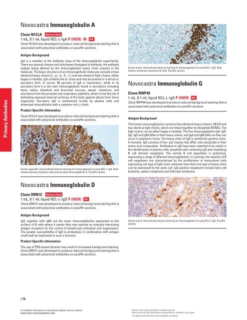
Human tonsil: immunohistochemical staining for Immunoglobulin G using NCL-L-IgG. Paraffin<br />
section.<br />
Products in this catalog are subject to regulatory approval.<br />
Please consult your Leica Microsystems representative for availability in your region.<br />
* For Research Use Only. Not for use in diagnostic procedures.


