

QF0159 Marketing Release Record
QF0159 Marketing Release Record
QF0159 Marketing Release Record

You also want an ePaper? Increase the reach of your titles
YUMPU automatically turns print PDFs into web optimized ePapers that Google loves.
Novocastra Peripherin<br />
Clone PJM50<br />
1 mL lyophilized NCL-PERIPH F P (HIER) W<br />
Peripherin is a 57 kD type III intermediate filament protein that is expressed<br />
in peripheral neurons, including enteric ganglion cells. Peripherin is<br />
expressed in the developing peripheral nervous system and is highly<br />
enriched in neuronal derivatives of the neural crest. The expression or<br />
absence of peripherin may be used to demonstrate abnormalities of the<br />
enteric nervous system. The assessment of the density of ganglion cells is of<br />
importance in Hirschsprung's disease (HD)-related disorders. Peripherin is<br />
also reported to be expressed in neural crest derived tumors such as<br />
neuroblastomas and ganglioneuroblastomas.<br />
Human small bowel: immunohistochemical staining for peripherin using NCL-PERIPH.<br />
Note intense cytoplasmic staining of enteric ganglion cells and neural elements.<br />
Paraffin section.<br />
Novocastra PETA-3 (CD151)<br />
Clone RLM30<br />
1 mL lyophilized NCL-CD151 P (HIER)<br />
See also CD151 (PETA-3) on page 46.<br />
Novocastra P-glycoprotein (CD243)<br />
Clone 5B12<br />
1 mL lyophilized NCL-PGLYm F P (HIER)<br />
Antigen Background<br />
RUO*<br />
The resistance of tumor cells to cytotoxic chemotherapeutic drugs is a major<br />
problem in the treatment of cancer. Studies have linked the presence of a 170<br />
to 180 kD cell membrane protein, P-glycoprotein, with resistance to a wide<br />
range of lipophilic chemotherapeutic drugs, a phenomenon known as<br />
multidrug resistance. P-glycoprotein is reported to be expressed in transporting<br />
epithelia of several normal tissues, including liver, kidney, colon,<br />
adrenal and brain.<br />
IVD<br />
RUO*<br />
Novocastra Placental Alkaline<br />
Phosphatase<br />
Clone 8A9<br />
1 mL, 0.1 mL lyophilized NCL-PLAP-8A9 F P (HIER)<br />
1 mL liquid NCL-L-PLAP-8A9 F P (HIER) IVD<br />
7 mL ready-to-use RTU-PLAP-8A9 F P (HIER) IVD<br />
7 mL BOND ready-to-use PA0161 P (HIER) IVD<br />
Antigen Background<br />
Placental alkaline phosphatase (PLAP) is a membrane-associated<br />
sialoglycoprotein enzyme normally present at high concentration in syncytiotrophoblasts<br />
within the placenta during the third trimester of gestation.<br />
The expression of PLAP was originally thought to be restricted to term<br />
placenta but a human PLAP-like variant has been described which shares<br />
more than 85 percent homology with PLAP itself. This high degree of<br />
homology between PLAP and PLAP-like enzyme together with cross-reacting<br />
antibodies has led to some confusion of the distribution of PLAP and PLAPlike<br />
enzyme in various tissues. PLAP is reported to be expressed only in<br />
normal term placenta, endocervix and fallopian tube and also in ovarian and<br />
proximal gastrointestinal tumors. PLAP expression is rare in malignant germ<br />
cell tumors. PLAP-like enzyme is reported to be predominantly found in<br />
normal fetal and neonatal testis, and in thymus. It is also commonly expressed<br />
in germ cell tumors and more recently described in seminomas.<br />
Product Specific Information<br />
Reports indicate that clone 8A9 stains seminomas and placenta indicating a<br />
specificity for both PLAP and PLAP-like enzyme.<br />
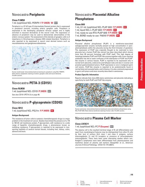
Human seminoma: immunohistochemical staining for placental alkaline phosphatase using<br />
NCL-PLAP-8A9. Note intense membrane staining of tumor cells. Paraffin section.<br />
Novocastra Plasma Cell Marker<br />
Clone LIV3G11<br />
1 mL lyophilized NCL-PC P (Enzyme)<br />
RUO*<br />
The plasma cell is the resultant terminal stage of B cell differentiation and<br />
apart from morphological features may be distinguished from other B cells<br />
by their lack of surface HLA class I and class II antigens, surface<br />
immunoglobulin, Fc and C3 receptors or presence of intracytoplasmic<br />
immunoglobulin. There have been some interesting studies where non-<br />
Hodgkin’s lymphomas have been differentiated from plasmacytomas and<br />
myelomas based on plasma cell markers (Seegmiller et al., American<br />
Journal of Clinical Pathology. 127(2):172-174 (2007)).<br />
F Frozen I Immunofluorescence E Electron microscopy<br />
P Paraffin C Flow cytometry O Other applications<br />
W Western blotting<br />
IVD<br />
/95<br />
Primary Antibodies


