

QF0159 Marketing Release Record
QF0159 Marketing Release Record
QF0159 Marketing Release Record

Create successful ePaper yourself
Turn your PDF publications into a flip-book with our unique Google optimized e-Paper software.
Novocastra Lambda Light Chain<br />
Clone SHL53 Reference Range<br />
1 mL, 0.1 mL liquid NCL-L-LAM-578 P (HIER)<br />
7 mL BOND ready-to-use PA0570 P (HIER)<br />
Clone HP-6054<br />
1 mL lyophilized NCL-LAM ASR<br />
Analyte Specific Reagent. Analytical and performance characteristics are<br />
not established.<br />
Novocastra Lamin<br />
Clone 636<br />
1 mL lyophilized lamin A/C NCL-LAM-A/C F P (HIER) W<br />
Antigen Background<br />
The nuclear lamina is a karyoskeletal structure composed of intermediate<br />
filament type proteins called lamins. It underlies the inner nuclear membrane<br />
and confers mechanical stability to the nuclear envelope. The human lamina<br />
consists of four major types of lamin, namely A, B1, B2 and C. The loss of lamin<br />
A expression has been reported to occur in small cell lung cancers.<br />
Product Specific Information<br />
NCL-LAM-A/C reacts with lamins A and C in human, cow and pig tissues.<br />
Novocastra Laminin<br />
Clone LAM-89<br />
0.5 mL lyophilized NCL-LAMININ F P (Enzyme)<br />
Laminin is a large (850 kD) disulfide-bonded heterotrimer, cross-shaped,<br />
glycoprotein which is organized within the meshwork of basement<br />
membranes such as those associated with epithelia, surrounding blood<br />
vessels, nerves and underlying pial sheaths of the brain. It is reported to be<br />
expressed in the extracellular matrix in sites other than basement<br />
membranes during early stages of development and is localized to specific<br />
types of neurons in the central nervous system during both embryonic and<br />
adult development. Laminin interacts with receptors on cell surfaces, an<br />
interaction which results in changes in the behavior of cells such as<br />
attachment to a substrate, migration and neurite outgrowth during<br />
embryonic development and regeneration.<br />
Human placenta: immunohistochemical staining for laminin using NCL-LAMININ. Note staining<br />
of basement membranes of blood vessels. Paraffin section.<br />
IVD<br />
IVD<br />
IVD<br />
IVD<br />
Novocastra Langerin<br />
Clone 12D6<br />
1 mL lyophilized NCL-LANGERIN P (HIER)<br />
Antigen Background<br />
Langerin is a type II transmembrane C-type lectin which has mannosebinding<br />
specificity. It is a 40 kD protein restricted to Langerhans cells that is<br />
involved in the internalization of cell surface material in these immature<br />
dendritic cells. Dendritic cells are antigen-presenting cells that are required<br />
for initiation of a specific T cell-driven immune response. These cells are<br />
found in nonlymphoid tissue as immature cells whose primary function is to<br />
capture antigen through specialized surface membrane endocytic structures<br />
or through macropinocytosis. The dendritic cells migrate to secondary<br />
lymphoid tissue and mature into efficient antigen presenting cells. A part of<br />
the maturation process includes the loss of adhesion receptors such as Ecadherin<br />
and the disappearance of Birbeck granules. Although Langerin is<br />
reported to be located on the cell surface, it can be rapidly internalized<br />
following ligand capture into Birbeck granules. In fact, Langerin is a potent<br />
inducer of membrane superimposition and zippering leading to Birbeck<br />
granule formation. In reports it has been suggested that the induction of<br />
Birbeck granules is a consequence of the antigen-capture function of<br />
Langerin allowing passage into these organelles and providing access to a<br />
non-classical antigen processing pathway.<br />
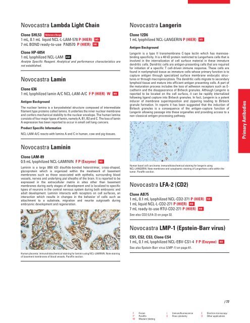
Human basal cell carcinoma: immunohistochemical staining for langerin using<br />
NCL-LANGERIN. Note membrane and cytoplasmic staining of Langerhans cells within the<br />
tumor. Paraffin section.<br />
Novocastra LFA-2 (CD2)<br />
Clone AB75<br />
1 mL, 0.1 mL lyophilized NCL-CD2-271 P (HIER)<br />
1 mL liquid NCL-L-CD2-271 P (HIER) IVD<br />
7 mL ready-to-use RTU-CD2-271 P (HIER) IVD<br />
See also CD2 (LFA-2) on page 32.<br />
Novocastra LMP-1 (Epstein-Barr virus)<br />
CS1, CS2, CS3, Clone CS4<br />
1 mL, 0.1 mL lyophilized NCL-EBV-CS1-4 F P (Enzyme)<br />
See also Epstein-Barr virus (LMP-1) on page 61.<br />
F Frozen I Immunofluorescence E Electron microscopy<br />
P Paraffin C Flow cytometry O Other applications<br />
W Western blotting<br />
IVD<br />
IVD<br />
IVD<br />
/77<br />
Primary Antibodies


