

QF0159 Marketing Release Record
QF0159 Marketing Release Record
QF0159 Marketing Release Record

Create successful ePaper yourself
Turn your PDF publications into a flip-book with our unique Google optimized e-Paper software.
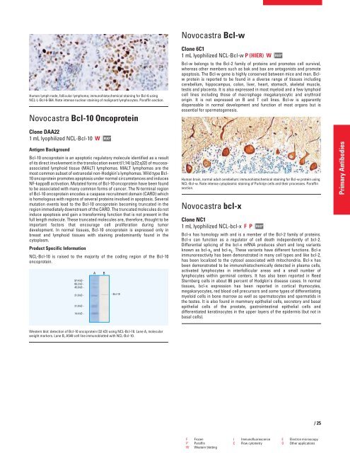
Human lymph node, follicular lymphoma; immunohistochemical staining for Bcl-6 using<br />
NCL-L-Bcl-6-564. Note intense nuclear staining of malignant lymphocytes. Paraffin section.<br />
Novocastra Bcl-10 Oncoprotein<br />
Clone DAA22<br />
1 mL lyophilized NCL-Bcl-10 W<br />
Antigen Background<br />
RUO*<br />
Bcl-10 oncoprotein is an apoptotic regulatory molecule identified as a result<br />
of its direct involvement in the translocation event t(1;14) (p22;q32) of mucosaassociated<br />
lymphoid tissue (MALT) lymphomas. MALT lymphomas are the<br />
most common subset of extranodal non-Hodgkin’s lymphomas. Wild type Bcl-<br />
10 oncoprotein promotes apoptosis under normal circumstances and induces<br />
NF-kappaB activation. Mutated forms of Bcl-10 oncoprotein have been found<br />
to be associated with many common forms of cancer. The N-terminal region<br />
of Bcl-10 oncoprotein encodes a caspase recruitment domain (CARD) which<br />
is homologous with regions of several proteins involved in apoptosis. Several<br />
mutation events lead to the Bcl-10 oncoprotein becoming truncated in the<br />
region immediately downstream of the CARD. The truncated molecules do not<br />
induce apoptosis and gain a transforming function that is not present in the<br />
full length molecule. These truncated molecules are, therefore, thought to be<br />
important factors that encourage cell proliferation during tumor<br />
development. In normal tissues, Bcl-10 oncoprotein is expressed only in<br />
breast and lymphoid tissues with staining predominantly found in the<br />
cytoplasm.<br />
Product Specific Information<br />
NCL-Bcl-10 is raised to the majority of the coding region of the Bcl-10<br />
oncoprotein.<br />
Western blot: detection of Bcl-10 oncoprotein (32 kD) using NCL-Bcl-10. Lane A, molecular<br />
weight markers. Lane B, A549 cell line immunoblotted with NCL-Bcl-10.<br />
Novocastra Bcl-w<br />
Clone 6C1<br />
1 mL lyophilized NCL-Bcl-w P (HIER) W<br />
Bcl-w belongs to the Bcl-2 family of proteins and promotes cell survival,<br />
whereas other members such as bak and bax are antagonists and promote<br />
apoptosis. The Bcl-w gene is highly conserved between mice and man. Bclw<br />
protein is reported to be found in a diverse range of tissues including<br />
cerebellum, hippocampus, colon, liver, heart, stomach, skeletal muscle,<br />
testis and placenta. It is also expressed in most myeloid and a few lymphoid<br />
cell lines including those of macrophage megakaryocytic and erythroid<br />
origin. It is not expressed on B and T cell lines. Bcl-w is apparently<br />
dispensable in normal development and function of most organs but is<br />
essential for spermatogenesis.<br />
Human brain, normal adult cerebellum: immunohistochemical staining for Bcl-w protein using<br />
NCL-Bcl-w. Note intense cytoplasmic staining of Purkinje cells and their processes. Paraffin<br />
section.<br />
Novocastra bcl-x<br />
Clone NC1<br />
1 mL lyophilized NCL-bcl-x FP<br />
RUO*<br />
RUO*<br />
Bcl-x has homology with and is a member of the Bcl-2 family of proteins.<br />
Bcl-x can function as a regulator of cell death independently of bcl-2.<br />
Differential splicing of the bcl-x mRNA produces short and long variants<br />
known as bcl-x s and bcl-x L . These variants have different functions. Bcl-x<br />
immunoreactivity has been demonstrated in many cell types and like bcl-2,<br />
has been localized to the cytosol associated with mitochondria. Bcl-x has<br />
been demonstrated to be immunohistochemically detected in plasma cells,<br />
activated lymphocytes in interfollicular areas and a small number of<br />
lymphocytes within germinal centers. It has also been reported in Reed<br />
Sternberg cells in about 86 percent of Hodgkin's disease cases. In normal<br />
tissues, bcl-x expression has been reported in cortical thymocytes,<br />
megakaryocytes, red blood cell precursors and some types of differentiating<br />
myeloid cells in bone marrow as well as spermatocytes and spermatids in<br />
the testes. It is also found in mammary epithelial cells, secretory and basal<br />
epithelial cells of the prostate, gastrointestinal epithelial cells and<br />
differentiated keratinocytes in the upper layers of the epidermis (but not in<br />
basal cells).<br />
F Frozen I Immunofluorescence E Electron microscopy<br />
P Paraffin C Flow cytometry O Other applications<br />
W Western blotting<br />
/25<br />
Primary Antibodies


