

QF0159 Marketing Release Record
QF0159 Marketing Release Record
QF0159 Marketing Release Record

Create successful ePaper yourself
Turn your PDF publications into a flip-book with our unique Google optimized e-Paper software.
Primary Antibodies<br />
Novocastra Dystrophin Antibodies<br />
Clone Dy4/6D3<br />
2.5 mL, 1 mL lyophilized Dystrophin (Rod Domain)<br />
NCL-DYS1 FWE IVD<br />
Clone Dy8/6C5<br />
2.5 mL, 1 mL lyophilized Dystrophin (C-terminus)<br />
NCL-DYS2 FWE IVD<br />
Clone Dy10/12B2<br />
2.5 mL, 1 mL lyophilized Dystrophin (N-terminus)<br />
NCL-DYS3 FWE IVD<br />
Clone 13H6<br />
1 mL lyophilized Dystrophin (C-terminus)<br />
NCL-DYSA P (HIER) RUO*<br />
Clone 34C5<br />
1 mL lyophilized Dystrophin (N-terminus)<br />
NCL-DYSB P (HIER) RUO*<br />
Antigen Background<br />
Duchenne Muscular dystrophy (DMD) is the most severe of the muscular<br />
dystrophies resulting in progressive muscular wasting and death. Dystrophin<br />
is the 427 kD protein product of the DMD/BMD gene located on the X<br />
chromosome at position Xp2. Western blotting and immunohisto-chemistry<br />
are the two established methods for the detection of abnormalities of<br />
dystrophin expression in muscle samples.<br />
Product Specific Information<br />
NCL-DYS1, NCL-DYS2 and NCL-DYS3 map within amino acids 1181-1388,<br />
3669-3685 and 321-494, respectively, on the dystrophin molecule. The<br />
immunolabeling patterns for NCL-DYS1, NCL-DYS2 and NCL-DYS3 are similar.<br />
NCL-DYSA is raised to a region of the dystrophin molecule, upstream from the<br />
C-terminal region and NCL-DYSB is raised to a region of the N-terminus of the<br />
dystrophin molecule. NCL-DYSA and NCL-DYSB will be of particular interest<br />
in the investigation of archived formalin-fixed, paraffin-embedded material.<br />
Labeling with an antibody to beta-spectrin, to monitor membrane integrity, is<br />
an essential immunohistochemical control.<br />
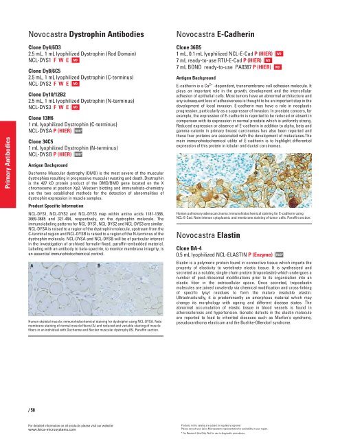
Human skeletal muscle: immunohistochemical staining for dystrophin using NCL-DYSA. Note<br />
membrane staining of normal muscle fibers (A) and reduced and variable staining of muscle<br />
fibers in an individual with Duchenne and Becker muscular dystrophy (B). Paraffin section.<br />
/58<br />
For detailed information on all products please visit our website:<br />
www.leica-microsystems.com<br />
Novocastra E-Cadherin<br />
Clone 36B5<br />
1 mL, 0.1 mL lyophilized NCL-E-Cad P (HIER)<br />
7 mL ready-to-use RTU-E-Cad P (HIER) IVD<br />
7 mL BOND ready-to-use PA0387 P (HIER)<br />
Antigen Background<br />
E-cadherin is a Ca 2+ -dependent, transmembrane cell adhesion molecule. It<br />
plays an important role in the growth, development and the intercellular<br />
adhesion of epithelial cells. Most tumors have an abnormal architecture and<br />
any subsequent loss of adhesiveness is thought to be an important step in the<br />
development of local invasion. E-cadherin may have a role in neoplastic<br />
progression, particularly as a suppressor of invasion. In prostate cancers, for<br />
example, the expression of E-cadherin is reported to be reduced or absent in<br />
comparison with its expression in normal prostate which is uniformly strong.<br />
Reduced expression or absence of E-cadherin in addition to alpha, beta and<br />
gamma-catenin in primary breast carcinomas has also been reported and<br />
these four proteins are associated with the development of metastases.The<br />
main immunohistochemical utility of E-cadherin is to highlight differential<br />
expression of this protein in lobular and ductal carcinomas.<br />
Human pulmonary adenocarcinoma: immunohistochemical staining for E-cadherin using<br />
NCL-E-Cad. Note intense cytoplasmic and membrane staining of tumor cells. Paraffin section.<br />
Novocastra Elastin<br />
Clone BA-4<br />
0.5 mL lyophilized NCL-ELASTIN P (Enzyme)<br />
Elastin is a polymeric protein found in connective tissue which imparts the<br />
property of elasticity to vertebrate elastic tissue. It is synthesized and<br />
secreted as a soluble, single-chain protein (tropoelastin) which undergoes a<br />
number of post-ribosomal modifications prior to its organization into an<br />
elastic fiber in the extracellular space. Once secreted, tropoelastin<br />
molecules are joined covalently via chemical modification and cross-linking<br />
of specific lysyl residues to form the mature insoluble elastin.<br />
Ultrastructurally, it is predominantly an amorphous material which may<br />
change its morphology with ageing and different disease states. The<br />
abnormal accumulation of elastic tissue in blood vessels is found in<br />
atherosclerosis and hypertension. Genetic defects in the elastin molecule<br />
are reported to lead to inherited diseases such as Marfan's syndrome,<br />
pseudoxanthoma elasticum and the Bushke-Ollendorf syndrome.<br />
Products in this catalog are subject to regulatory approval.<br />
Please consult your Leica Microsystems representative for availability in your region.<br />
* For Research Use Only. Not for use in diagnostic procedures.<br />
IVD<br />
IVD<br />
RUO*


