

QF0159 Marketing Release Record
QF0159 Marketing Release Record
QF0159 Marketing Release Record

Create successful ePaper yourself
Turn your PDF publications into a flip-book with our unique Google optimized e-Paper software.
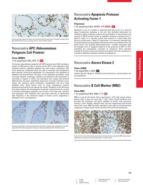
Human prostatic adenocarcinoma: immunohistochemical staining for androgen receptor using<br />
NCL-AR-318. Note nuclear staining of tumor cells. Paraffin section.<br />
Novocastra APC (Adenomatous<br />
Polyposis Coli Protein)<br />
Clone EMM43<br />
1 mL lyophilized NCL-APC P<br />
The human adenomatous polyposis coli (APC) gene at locus 5q21 encodes a<br />
protein of 2,843 amino acids. A precise role for APC in the regulation of the<br />
wnt/beta-cateninin signalling pathway has been clearly recognized. APC<br />
forms molecular complexes which are able to eliminate intra-cytoplasmic<br />
beta-catening, inducing its degradation. It is expressed in the cytoplasm of<br />
epithelial and mesenchymal cell types. In the epithelium of bladder, small<br />
and large intestine, esophagus, stomach and epidermis, APC expression is<br />
restricted to regions in which cell replication has ceased and terminal<br />
differentiation has been established. Expression has been reported in lung,<br />
kidney and mammary gland endothelial, myoepithelial and duct lining<br />
epithelial cells. Some tissues such as ovary, myometrium, thyroid,<br />
parathyroid and tonsil do not express the protein. Mutations of the APC gene<br />
have been linked to the development of sporadic colorectal tumors, as well<br />
as familial adenomatous polyposis and cancers of the pancreas, stomach<br />
and esophagus. APC mutations have also been observed at significantly<br />
high frequency in the advanced stages of breast cancer suggesting a<br />
biological role in carcinogenesis.<br />
Human small intestine: immunohistochemical staining for adenomatous polyposis coli protein<br />
using NCL-APC. Note cytoplasmic staining of intestinal epithelial cells. Paraffin section.<br />
IVD<br />
Novocastra Apoptosis Protease<br />
Activating Factor 1<br />
Polyclonal<br />
1 mL lyophilized NCL-APAF1 F P (HIER)<br />
Apoptosis is one of a number of responses that may occur as a result of<br />
signal transduction pathways in the cell. One identified mechanism for<br />
initiating caspase activation requires the participation of mitochondria and<br />
involves a 130 kD protein known as apoptosis protease activating factor-1<br />
(Apaf-1). Apaf-1 is a cytosolic protein that remains in a latent state until<br />
bound to cytochrome c (Apaf-2). Cytochrome c is commonly released from<br />
the mitochondria during apoptosis induced by many, but probably not all cell<br />
death stimuli. The resulting Apaf1/cytochrome c complex associates with<br />
the zymogen form of caspase-9 (Apaf-3) in the presence of dATP or ATP,<br />
promoting the autocatalytic activation of caspase-9. Once activated<br />
caspase-9 can then cleave and activate procaspase-3 directly, resulting in a<br />
cascade of additional caspase activation and apoptosis.<br />
Novocastra Aurora Kinase 2<br />
Clone JLM28<br />
1 mL liquid NCL-L-AK2 ASR<br />
Analyte Specific Reagent. Analytical and performance characteristics are<br />
not established.<br />
Novocastra B Cell Marker (MB2)<br />
Clone MB2<br />
1 mL lyophilized NCL-MB2 FP<br />
RUO*<br />
MB2 is a pan B cell marker that is expressed in all B cells except mature<br />
plasma cells. It does not react with T cells. These include epidermis (but<br />
excludes the squamous cell layer), epithelia of breast, lung, pancreas,<br />
stomach, colon, bladder, fallopian tube and also hepatocytes and stromal<br />
cells of the ovary. MB2 has been reported to react with an uncharacterized<br />
cytoplasmic antigen found in both normal B cells and B cell lymphomas.<br />
Human tonsil: immunohistochemical staining for B lymphocytes using NCL-MB2. Note intense<br />
cytoplasmic staining of normal B lymphocytes. Paraffin section.<br />
F Frozen I Immunofluorescence E Electron microscopy<br />
P Paraffin C Flow cytometry O Other applications<br />
W Western blotting<br />
IVD<br />
/23<br />
Primary Antibodies


