

QF0159 Marketing Release Record
QF0159 Marketing Release Record
QF0159 Marketing Release Record

You also want an ePaper? Increase the reach of your titles
YUMPU automatically turns print PDFs into web optimized ePapers that Google loves.
Primary Antibodies<br />
Novocastra MDM2 Protein<br />
Clone 1B10<br />
1 mL lyophilized NCL-MDM2 F P (HIER)<br />
Antigen Background<br />
The human phosphoprotein homolog of the murine double minute 2 (MDM2)<br />
gene, with a molecular weight of 90 kD (p90), forms a complex with both<br />
mutant and wild type p53 protein. The MDM2 gene product interacts with p53<br />
protein inhibiting p53-mediated transactivation. Overexpression of MDM2<br />
overcomes wild type p53 mediated suppression of transformed cell growth.<br />
MDM2 amplification is reported to be observed in some soft tissue sarcomas,<br />
osteosarcomas and high grade malignant gliomas.<br />
Product Specific Information<br />
NCL-MDM2 reacts with the human homolog of MDM2.<br />
Human breast carcinoma: immunohistochemical staining for MDM2 protein using NCL-MDM2.<br />
Note nuclear staining of tumor cells. Paraffin sectiion.<br />
Novocastra Melan A<br />
Clone A103 Reference Range<br />
1 mL, 0.1 mL lyophilized NCL-MelanA F P (HIER) W<br />
1 mL liquid NCL-L-MelanA F P (HIER) W IVD<br />
7 mL ready-to-use RTU-MelanA F P (HIER) IVD<br />
7 mL BOND ready-to-use PA0233 P (HIER) IVD<br />
Antigen Background<br />
Melan A, a product of the MART-1 gene, is a melanocyte differentiation<br />
marker recognized by autologous cytotoxic T lymphocytes. Other melanomaassociated<br />
markers recognized by autologous cytotoxic T cells are reported<br />
to include MAGE-1, MAGE-3, tyrosinase, gp100, gp75, BAGE-1 and GAGE-1.<br />
The analysis of these different molecules and their expression in individual<br />
melanomas may be of help in the study of their particular molecular roles in<br />
melanocyte differentiation and tumorigenesis.<br />
Human melanoma: immunohistochemical staining for melan A using NCL-MelanA.<br />
Note cytoplasmic staining of melanoma cells. Paraffin section.<br />
/80<br />
For detailed information on all products please visit our website:<br />
www.leica-microsystems.com<br />
IVD<br />
IVD<br />
Novocastra Melanoma Marker (CD63)<br />
Clone NKI/C3<br />
1 mL lyophilized NCL-CD63 FP<br />
See also CD63 (Melanoma Marker) on page 42.<br />
Novocastra Melanoma Marker (HMB45)<br />
Clone HMB45 Reference Range<br />
1 mL, 0.1mL liquid NCL-HMB45 F P (Enzyme)<br />
7 mL BOND ready-to-use PA0027 P (Enzyme)<br />
See also HMB45 (Melanoma Marker) on page 71.<br />
Novocastra Merosin Laminin Alpha 2<br />
Chain<br />
Clone Mer3/22B2<br />
1 mL, 0.1mL lyophilized NCL-MEROSIN F<br />
Antigen Background<br />
The dystrophin-glycoprotein complex is localized to the muscle membrane.<br />
Several members of this complex are reported to be implicated in muscular<br />
dystrophy. Dystrophin expression is altered in Duchenne and Becker<br />
muscular dystrophy and four types of limb girdle muscular dystrophy are<br />
caused by mutations in the genes for alpha, beta, gamma and deltasarcoglycan.<br />
An extracellular member of this complex is alpha-dystroglycan<br />
and linked to this, in the extracellular matrix, is laminin. The muscle specific<br />
form of laminin, merosin, is composed of three chains: alpha 2, beta 1 and<br />
gamma 1. Mutations in the chromosome 6 encoded gene for the laminin alpha<br />
2 chain of merosin are responsible for a form of congenital muscular<br />
dystrophy (CMD). Merosin negative CMD is characterized by a severe clinical<br />
phenotype and is associated with white matter changes on brain imaging.<br />
Product Specific Information<br />
NCL-MEROSIN reacts with the 300 kD fragment of merosin (Sewry et al.<br />
Muscle and Nerve Supplement. 7, S109: (1998)) labeling with an antibody to<br />
beta-spectrin to monitor membrane integrity, is an essential immunohistochemical<br />
control.<br />
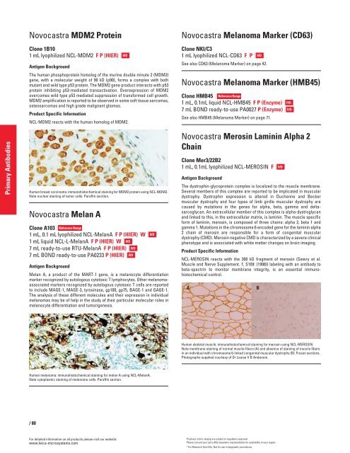
A B<br />
Human skeletal muscle: immunohistochemical staining for merosin using NCL-MEROSIN.<br />
Note membrane staining of normal muscle fibers (A) and absence of staining of muscle fibers<br />
in an individual with chromosome 6-linked congenital muscular dystrophy (B). Frozen sections.<br />
Photographs supplied courtesy of Dr Louise V B Anderson.<br />
IVD<br />
Products in this catalog are subject to regulatory approval.<br />
Please consult your Leica Microsystems representative for availability in your region.<br />
* For Research Use Only. Not for use in diagnostic procedures.<br />
IVD<br />
IVD<br />
IVD


