

QF0159 Marketing Release Record
QF0159 Marketing Release Record
QF0159 Marketing Release Record

Create successful ePaper yourself
Turn your PDF publications into a flip-book with our unique Google optimized e-Paper software.
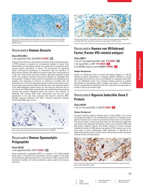
Human tonsil: immunohistochemical staining for human neutrophil defensins using NCL-<br />
DEFENSIN. Note intense granular cytoplasmic and extracellular staining of neutrophils.<br />
Paraffin section.<br />
Novocastra Human Securin<br />
Clone DCS-280.2<br />
1 mL lyophilized NCL-SECURIN P (HIER)<br />
Human securin (hsecurin), also known as pituitary tumor-transforming gene-<br />
1 (PTTG) product, is required for chromosomal stability in human cells.<br />
Abnormalities of chromosome number are reported to be amongst the most<br />
common genetic aberrations in cancer. The mechanisms for regulating<br />
mitotic chromosome transmission in mammalian cells are, therefore, of<br />
great interest. Human cells without an hsecurin gene lose chromosomes at<br />
a high rate. These losses have been linked to abnormal anaphases during<br />
which cells undergo repeated unsuccessful attempts to segregate their<br />
chromosomes. Therefore, human securin is essential for the maintenance of<br />
euploidy. The expression of hsecurin is reported to correlate with cell<br />
proliferation in a cell cycle-dependent manner in both normal tissues and in<br />
several tumor types. hsecurin specifically binds to Ku, the regulatory subunit<br />
of the DNA-dependent protein kinase. Ku and hsecurin associate both in<br />
vitro and in vivo. DNA double-strand breaks prevent Ku/hsecurin association<br />
showing that genome damaging events can result in the induction of<br />
pathways that activate DNA repair mechanisms and halt cell cycle<br />
progression. It has also been proposed that hsecurin connects DNAdamage<br />
response pathways with sister chromatid separation delaying<br />
mitosis while DNA repair occurs.<br />
Human tonsil: immunohistochemical staining for human securin using NCL-SECURIN.<br />
Note nuclear staining of proliferating cells. Paraffin section, nickel DAB.<br />
Novocastra Human Spasmolytic<br />
Polypeptide<br />
Clone GE16C<br />
1 mL lyophilized NCL-HSP P (HIER)<br />
RUO*<br />
RUO*<br />
Human spasmolytic polypeptide (HSP) is a member of the trefoil peptide<br />
family which is reported to be expressed in discrete regions of the body,<br />
most notably the gastrointestinal tract. In the stomach, HSP is reported to be<br />
localized to foveolar and surface epithelium, pyloric glands and mucous<br />
neck cells.<br />
Normal human stomach: immunohistochemical staining of human spasmolytic polypeptide<br />
using NCL-HSP. Note cytoplasmic staining of cardiac glands. Paraffin section.<br />
Novocastra Human von Willebrand<br />
Factor (Factor VIII-related antigen)<br />
Clone 36B11<br />
1 mL, 0.1 mL lyophilized NCL-vWF F P (HIER)<br />
1 mL liquid NCL-L-vWF F P (HIER) IVD<br />
7 mL BOND ready-to-use PA0400 P (HIER)<br />
Antigen Background<br />
Human von Willebrand factor (or factor VIII-related antigen) is a 270 kD<br />
multimeric plasma glycoprotein. It mediates platelet adhesion to injured<br />
vessel walls and serves as a carrier and stabilizer for coagulation factor VIII.<br />
The von Willebrand factor has functional binding domains to platelet<br />
glycoprotein Ib, glycoprotein Ib/IIIa, collagen and heparin. von Willebrand<br />
factor is synthesized by endothelial cells and is reported to be expressed in a<br />
number of tumors of vascular origin.<br />
Novocastra Hypoxia Inducible Gene 2<br />
Protein<br />
Clone HX34Y<br />
1 mL, 0.1 mL liquid NCL-L-HIG2 P (HIER)<br />
Antigen Background<br />
The gene encoding hypoxia-inducible gene 2 protein (HIG2) is one of the<br />
transcriptional targets for the activated beta-catenin/Tcf-4 complex and its<br />
product functions as an autocrine growth factor that enhances cell growth.<br />
This gene encodes a trans-membrane protein of 7 kD molecular weight that<br />
was found to be expressed exclusively in renal cell carcinomas (RCC) and<br />
fetal kidney. Reports indicate that ELISA analysis of clinical samples<br />
identified secretion of HIG2 protein into plasma of RCC patients even at an<br />
early stage of tumor development. HIG2 expression is reported to be<br />
expressed at higher levels in ovarian clear cell carcinomas when compared<br />
to those of clear cell renal tumors.<br />
IVD<br />
RUO*<br />
Human clear cell renal cell carcinoma: immunohistochemical staining for hypoxia inducible<br />
gene 2 protein using NCL-L-HIG2.<br />
F Frozen I Immunofluorescence E Electron microscopy<br />
P Paraffin C Flow cytometry O Other applications<br />
W Western blotting<br />
IVD<br />
/73<br />
Primary Antibodies


