

QF0159 Marketing Release Record
QF0159 Marketing Release Record
QF0159 Marketing Release Record

Create successful ePaper yourself
Turn your PDF publications into a flip-book with our unique Google optimized e-Paper software.
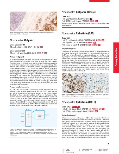
Human medullary thyroid carcinoma: immunohistochemical staining for calcitonin using<br />
NCL-L- CALCITONIN. Paraffin section.<br />
Novocastra Calpain<br />
Clone Calp3c/11B3<br />
2.5 mL lyophilized NCL-CALP-11B3 W<br />
Clone Calp3c/12A2<br />
2.5 mL, 1 mL lyophilized NCL-CALP-12A2 W<br />
Antigen Background<br />
RUO*<br />
RUO*<br />
At least seven forms of autosomal recessive muscular dystrophy (MD) have<br />
been included under the banner “limb girdle muscular dystrophy” (LGMD).<br />
These forms may be divided into two groups; those with abnormal expression<br />
of the dystrophin/glycoprotein complex and those in which labeling of the<br />
proteins in this complex is unaffected. Thus the sarcoglycanopathies (also<br />
known as LGMD types 2C, 2D, 2E and 2F) are caused by defects in the genes<br />
for gamma, alpha, beta and delta-sarcoglycan on chromosomes 13q12, 17q21,<br />
4q12 and 5q33, respectively. Among the dystrophies in which expression of<br />
the sarcoglycans is normal, the gene responsible for LGMD2A has been<br />
identified as the chromosome 15q15-encoded muscle-specific calciumactivated<br />
neutral protease, calpain 3. Calpain 3 enzyme is only stable in<br />
human muscle when homogenized in treatment buffer immediately after<br />
harvest. (Anderson LVB et al. American Journal of Pathology. 153(4): 1169-<br />
1179 (1998)), and in homogenates containing SDS and is therefore well suited<br />
for analysis by Western blot.<br />
Product Specific Information<br />
NCL-CALP-2C4 reacts with the full-size calpain 3 (94 kD) and an additional<br />
fragment (30 kD) in human skeletal muscle. NCL-CALP-12A2 reacts with fullsize<br />
protein plus apparent degradation products at approximately 60 kD.<br />
Specificity of these antibodies has been confirmed by the loss of all these<br />
bands in samples with null gene mutations. NCL-CALP-11B3 reacts with<br />
calpain 3 bands at 94 and 60 kD, pre- and post-autolyzed forms of the<br />
ubiquitous calpains 1 and 2 (� and m-calpain) staining a group of bands<br />
between 76 and 84 kD in human skeletal muscle (Anderson LVB et al.<br />
American Journal of Pathology. 153(4): 1169-1179, (1998)) Cross-reactivities in<br />
different animals and tissues are described (see reference).<br />
Western blot: analysis of human skeletal muscle showing detection of the calpain family of<br />
proteins. Lane A, calpain 3 bands at 94 and 30 kD detected with NCL-CALP-2C4. Lane B,<br />
calpains 1, 2 and 3 detected with NCL-CALP-11B3. Lane C, calpain 3 bands at 94 and<br />
approximately 60 kD detected with NCL-CALP-12A2. Photograph supplied courtesy of Dr Louise<br />
V B Anderson.<br />
Novocastra Calponin (Basic)<br />
Clone 26A11<br />
1 mL lyophilized NCL-CALPONIN-B ASR<br />
7 mL BOND ready-to-use PA0416 P (HIER) IVD<br />
Analyte Specific Reagent. Analytical and performance characteristics are<br />
not established.<br />
Novocastra Calretinin (5A5)<br />
Clone 5A5<br />
1 mL, 0.1 mL lyophilized NCL-CALRETININ P (HIER)<br />
1 mL liquid NCL-L-CALRETININ P (HIER) IVD<br />
7 mL ready-to-use RTU-CALRETININ P (HIER) IVD<br />
Antigen Background<br />
Calretinin is an intracellular calcium-binding protein of 31.5 kD belonging to<br />
the troponin C superfamily characterized by a structural motif described as<br />
the EF-hand domain. Calcium is an important moderator of a number of vital<br />
physiological processes, including neuronal excitability, axonal transport,<br />
synthesis and release of some neurotransmitters, membrane permeability<br />
and enzyme activity. Calretinin is found in the nervous system and thymus.<br />
Calretinin can also be demonstrated in normal and neoplastic mesothelial<br />
cells and has been reported to be a useful marker for the identification of<br />
malignant mesotheliomas of epithelial type to differentiate these from<br />
metastases of lung adenocarcinoma where antibodies to detect calretinin<br />
are used in within a panel and interpretation together with clinical data is<br />
undertaken by a qualified pathologist.<br />
Human mesothelioma: immunohistochemical staining for calretinin using NCL-L-CALRETININ.<br />
Note cytoplasmic staining of malignant cells. Paraffin section.<br />
Novocastra Calretinin (CAL6)<br />
Clone CAL6 Reference Range<br />
1 mL, 0.1 mL liquid NCL-L-CALRET-566 P (HIER) W<br />
7 mL BOND ready-to-use PA0346 P (HIER) IVD<br />
Antigen Background<br />
Calretinin is a calcium-binding protein of 29 kD that is a member of the family<br />
of so-called EF-hand proteins that also includes S-100 proteins. Calretinin is<br />
reported to be abundantly expressed in neurons. Outside the nervous system,<br />
calretinin is reported to be expressed in a range of cell types including<br />
mesothelial cells, steroid producing cell, (eg adrenal cortical cells, Leydig<br />
cells, ovarian theca interna cells as well as Sertoli cells, some<br />
neuroendocrine cells, eccrine sweat glands) and other cell types. The<br />
presence of calretinin is reported to be a useful marker primarily for<br />
differentiating malignant mesothelioma from carcinomas. Other reports<br />
indicate the detection of calretinin is helpful for the differential<br />
characterization of ovarian stroma tumors. Calretinin-positive cells have also<br />
been reported in the convoluted tubules of kidney with some antibodies.<br />
F Frozen I Immunofluorescence E Electron microscopy<br />
P Paraffin C Flow cytometry O Other applications<br />
W Western blotting<br />
IVD<br />
IVD<br />
/29<br />
Primary Antibodies


