

QF0159 Marketing Release Record
QF0159 Marketing Release Record
QF0159 Marketing Release Record

Create successful ePaper yourself
Turn your PDF publications into a flip-book with our unique Google optimized e-Paper software.
Primary Antibodies<br />
Novocastra CD81<br />
Clone 1D6<br />
1 mL lyophilized NCL-CD81 P (HIER)<br />
CD81 antigen, also known as TAPA-1, is a member of the TM4 superfamily<br />
and is predicted to have four transmembrane regions, short cytoplasmic<br />
N and C-termini and two extracellular regions. CD81 protein has a molecular<br />
weight of 26 kD and is expressed by most cell types. Of the hematopoietic<br />
cells, CD81 protein is reported to be expressed by B and T cells,<br />
macrophages, dendritic cells, NK cells and eosinophils but not by<br />
neutrophils, platelets or erythrocytes. The CD81 protein associates noncovalently<br />
with a number of other molecules eg CD19, CD21, MHC class<br />
I and II, CD20, CD37, CD53 and CD82 in B cells and CD4, CD8 and CD82 in<br />
T cells. It also associates with the integrins CD29/CD49c (VLA-3), CD29/<br />
CD49d (VLA-4) and CD29/CD49f (VLA-6) in several other cell types. No<br />
extracellular ligand has been identified for CD81 protein and its function<br />
remains unclear, although mouse CD81 protein plays a role in early T cell<br />
development. The human CD81 molecule has been reported to be involved in<br />
cell adhesion, motility, metastasis as well as cell activation and signal<br />
transduction.<br />
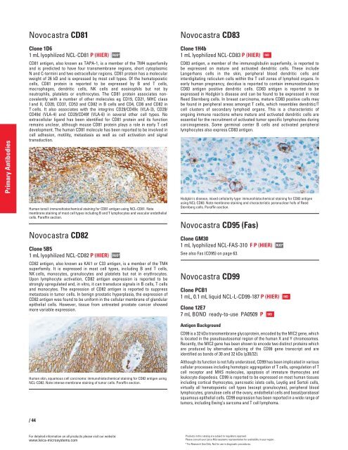
Human tonsil: immunohistochemical staining for CD81 antigen using NCL-CD81. Note<br />
membrane staining of most cell types including B and T lymphocytes and vascular endothelial<br />
cells. Paraffin section.<br />
Novocastra CD82<br />
Clone 5B5<br />
1 mL lyophilized NCL-CD82 P (HIER)<br />
/44<br />
RUO*<br />
RUO*<br />
CD82 antigen, also known as KAI1 or C33 antigen, is a member of the TM4<br />
superfamily. It is expressed in most cell types, including B and T cells,<br />
NK cells, monocytes, granulocytes and platelets but not in erythrocytes.<br />
Upon lymphocyte activation, CD82 antigen expression is reported to be<br />
strongly upregulated and, in vitro, it can transduce signals in B cells, T cells<br />
and monocytes. The expression of CD82 antigen is reported to suppress<br />
metastasis in tumor cells. In benign prostatic hyperplasia, the expression of<br />
CD82 antigen was found to be uniform in the cellular membrane of glandular<br />
epithelial cells. However, tissue from untreated prostate cancer showed<br />
more variable expression.<br />
Human skin, squamous cell carcinoma: immunohistochemical staining for CD82 antigen using<br />
NCL-CD82. Note intense membrane staining of tumor cells. Paraffin section.<br />
For detailed information on all products please visit our website:<br />
www.leica-microsystems.com<br />
Novocastra CD83<br />
Clone 1H4b<br />
1 mL lyophilized NCL-CD83 P (HIER)<br />
CD83 antigen, a member of the immunoglobulin superfamily, is reported to<br />
be expressed on mature and activated dendritic cells. These include<br />
Langerhans cells in the skin, peripheral blood dendritic cells and<br />
interdigitating reticulum cells within the T cell zones of lymphoid organs. In<br />
early human pregnancy, decidua is reported to contain immunostimulatory<br />
CD83 antigen positive dendritic cells. CD83 antigen is reported to be<br />
expressed in Hodgkin's disease and can be found to be expressed in most<br />
Reed Sternberg cells. In breast carcinoma, mature CD83 positive cells may<br />
be found in peripheral areas amongst T cells, which resembles dendritic/T<br />
cell clusters of secondary lymphoid organs. This is a characteristic of<br />
ongoing immune reactions where mature and activated dendritic cells are<br />
essential for the recruitment of activated tumor specific lymphocytes during<br />
carcinogenesis. Some germinal center B cells and activated peripheral<br />
lymphocytes also express CD83 antigen.<br />
Hodgkin's disease, mixed cellularity type: immunohistochemical staining for CD83 antigen<br />
using NCL-CD83. Note membrane staining and characteristic paranuclear hofs of Reed<br />
Sternberg cells. Paraffin section.<br />
Novocastra CD95 (Fas)<br />
Clone GM30<br />
1 mL lyophilized NCL-FAS-310 F P (HIER)<br />
See also Fas (CD95) on page 63.<br />
Novocastra CD99<br />
Clone PCB1<br />
1 mL, 0.1 mL liquid NCL-L-CD99-187 P (HIER)<br />
Clone 12E7<br />
7 mL BOND ready-to-use PA0509 P<br />
Antigen Background<br />
CD99 is a 32 kDa transmembrane glycoprotein, encoded by the MIC2 gene, which<br />
is located in the pseudoautosomal region of the human X and Y chromosomes.<br />
Recently, the MIC2 gene has been shown to encode two distinct proteins which<br />
are produced by alternative splicing of the CD99 gene transcript and are<br />
identified as bands of 30 and 32 kDa (p30/32).<br />
Although its function is not fully understood, CD99 has been implicated in various<br />
cellular processes including homotypic aggregation of T cells, upregulation of T<br />
cell receptor and MHS molecules, apoptosis of immature thymocytes and<br />
leukocyte diapedesis. CD99 is reported to be expressed on most human tissues<br />
including cortical thymocytes, pancreatic islets cells, Leydig and Sertoli cells,<br />
virtually all hematopoietic cell types (except granulocytes), peripheral blood<br />
lymphocytes, granulose cells of the ovary, endothelial cells and basal/parabasal<br />
squamous epithelial cells. CD99 expression has been reported in a wide range of<br />
tumors, including Ewing’s sarcoma and T cell lymphoma.<br />
IVD<br />
IVD<br />
Products in this catalog are subject to regulatory approval.<br />
Please consult your Leica Microsystems representative for availability in your region.<br />
* For Research Use Only. Not for use in diagnostic procedures.<br />
RUO*<br />
IVD


