

QF0159 Marketing Release Record
QF0159 Marketing Release Record
QF0159 Marketing Release Record

Create successful ePaper yourself
Turn your PDF publications into a flip-book with our unique Google optimized e-Paper software.
Product Specific Information<br />
Please note that methacarn fixation produces optimal staining.<br />
Human breast carcinoma: immunohistochemical staining for cyclin B1 using NCL-CYCLIN B1.<br />
Note cytoplasmic staining of tumor cells. Paraffin section.<br />
Novocastra Cyclin D1<br />
Clone P2D11F11 Reference Range<br />
1 mL, 0.1 mL lyophilized<br />
NCL-CYCLIN D1-GM P (HIER/Enzyme) W IVD<br />
1 mL liquid NCL-L-CYCLIN D1-GM P (HIER/Enzyme) W<br />
7 mL ready-to-use RTU-CYCLIN D1-GM P (HIER/Enzyme)<br />
Clone DCS-6<br />
1 mL lyophilized NCL-CYCLIN D1 F P (HIER/Enzyme) W<br />
Antigen Background<br />
The D-type cyclins are a family of proteins which function primarily by<br />
regulating the activity of cyclin dependent kinases in the G1 phase of the cell<br />
cycle. Cyclin D1, a protein of 36 kD, is also known as PRAD1 or bcl-1.<br />
Maximum expression of cyclin D1 occurs at a critical point in mid to late G1<br />
phase of the cell cycle. The cyclin D1 gene, located on 11q13 has been<br />
reported to be overexpressed in mantle cell lymphomas due to the<br />
chromosomal translocation t(11;18). It has also been observed in approximately<br />
30 percent of breast cancers.<br />
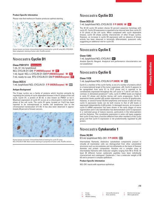
Human breast carcinoma: immunohistochemical staining for cyclin D1 using<br />
NCL-CYCLIN D1-GM. Note nuclear staining of a proportion of tumor cells. Paraffin section.<br />
IVD<br />
IVD<br />
RUO*<br />
Novocastra Cyclin D3<br />
Clone DCS-22<br />
1 mL lyophilized NCL-CYCLIN D3 F P (HIER) W<br />
The 34 kD cyclin D3 protein shares 53 percent sequence homology with<br />
cyclin D1. Cyclin D3 expression is reported to be induced later than cyclin D1<br />
in G1 phase of the cell cycle. When complexed with cyclin dependent<br />
kinases, cyclin D3 shows activity characteristic of other D-type cyclins.<br />
However, an increase in cyclin D3 expression with an absence of kinase<br />
activity has been observed in terminally differentiated, quiescent cells,<br />
suggesting an additional role for cyclin D3.<br />
Novocastra Cyclin E<br />
Clone 13A3<br />
1 mL lyophilized NCL-CYCLIN E ASR<br />
Analyte Specific Reagent. Analytical and performance characteristics are<br />
not established.<br />
Novocastra Cyclin G<br />
Clone 11C8<br />
1 mL lyophilized NCL-CYCLIN G P (HIER) W<br />
Cyclin G, a member of the cyclin family, is one of a number of proteins which<br />
is a transcriptional target of the tumor suppressor, p53. Cyclin G appears to<br />
be upregulated from early G1 to G1/S phase and is reported to be<br />
constitutively expressed throughout the cell cycle in T and B cell lines. In<br />
contrast, in stimulated peripheral T cells, cyclin G mRNA levels are highest<br />
in early G1 phase and decline during cell cycle progression. Cyclin G<br />
expression levels parallel p53 protein expression in murine B lymphocytes,<br />
however, in several human Burkitt lymphomas and tissues of p53 null mice,<br />
cyclin G expression levels can be both inverse to that of p53 levels or<br />
expressed independently of p53 protein. In damaged neurons, an increase in<br />
cyclin G mRNA expression has been shown in the early stages of nerve<br />
regeneration and in situ hybridization has demonstrated cyclin G expression<br />
in a restricted group of mature neurons, particularly in the telencephalon<br />
and the thalamus. This constitutive expression in some cell types suggests<br />
that cyclin G may have a function different from other members of the cyclin<br />
group and that cyclin G expression is not predominantly regulated by p53<br />
protein.<br />
Novocastra Cytokeratin 1<br />
Clone 34�B4<br />
0.5 mL lyophilized NCL-CK1 F P (HIER)<br />
Intermediate filaments, distinctive cytoskeletal components present in<br />
virtually all mammalian cells are distinguished from other cytoskeletal<br />
structures such as microtubules and microfilaments on the basis of filament<br />
diameter and protein composition. Keratins are a complex class of<br />
intermediate filaments with molecular weights ranging from 40 to 70 kD. At<br />
least 20 different human cytokeratin peptides have been individually<br />
characterized and catalogued. Cytokeratin 1 has a molecular weight of 68<br />
kD and is present in complex epithelium.<br />
Product Specific Information<br />
NCL-CK1 reacts with squamous epithelium.<br />
RUO*<br />
RUO*<br />
F Frozen I Immunofluorescence E Electron microscopy<br />
P Paraffin C Flow cytometry O Other applications<br />
W Western blotting<br />
IVD<br />
/51<br />
Primary Antibodies


