

QF0159 Marketing Release Record
QF0159 Marketing Release Record
QF0159 Marketing Release Record

You also want an ePaper? Increase the reach of your titles
YUMPU automatically turns print PDFs into web optimized ePapers that Google loves.
Novocastra Glial Fibrillary Acidic<br />
Protein<br />
Clone GA5<br />
1 mL lyophilized NCL-GFAP-GA5 FP IVD<br />
7 mL BOND ready-to-use PA0026 P (HIER)<br />
Antigen Background<br />
Glial fibrillary acidic protein (GFAP) is an intermediate filament protein of<br />
52 kD reported to be expressed in glial cells eg astrocytes and ependymal<br />
cells. In the peripheral nervous system, GFAP has been reported to be<br />
expressed in Schwann cells, enteric glial cells and satellite cells of human<br />
sensory ganglia and in neoplastic tissues GFAP has been reported to be<br />
expressed in astrocytomas and ependymomas.<br />
Product Specific Information<br />
When using NCL-GFAP-GA5 the heat induced epitope retrieval (HIER)<br />
technique may improve staining in some cases.<br />
Novocastra Glucagon<br />
Polyclonal<br />
0.5 mL lyophilized NCL-GLUCp FP IVD<br />
Polyclonal<br />
7 mL BOND ready-to-use PA0594 P (HIER)<br />
Antigen Background<br />
Glucagon expression has been reported in the endocrine cells of the<br />
pancreatic islets and also in the mucosa of small and large intestine.<br />
Pancreatic glucagon, a peptide of 29 amino acids, has biological activities<br />
including glycogenolysis, lipolysis, gluconeogenesis and ketogenesis. These<br />
effects are all antagonistic to insulin action and, therefore, lead to increased<br />
blood sugar levels. The majority of glucagonomas are reported to arise from<br />
the pancreas and produce pancreatic glucagon. These tumors are found<br />
chiefly in the main body or tail of the pancreas.<br />
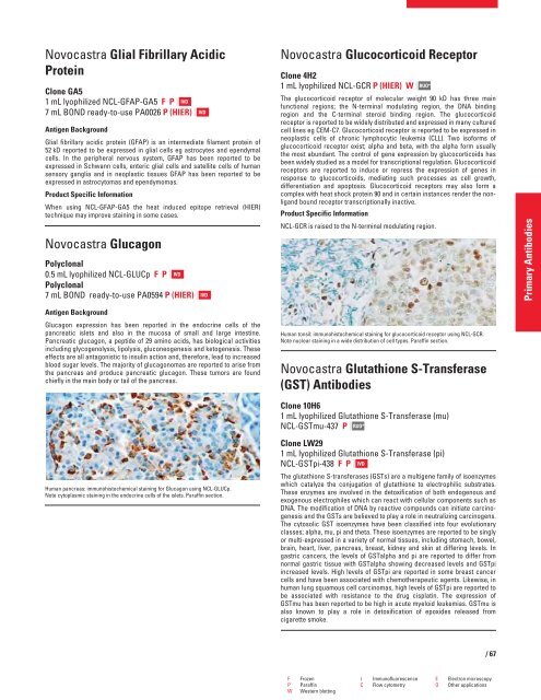
Human pancreas: immunohistochemical staining for Glucagon using NCL-GLUCp.<br />
Note cytoplasmic staining in the endocrine cells of the islets. Paraffin section.<br />
IVD<br />
IVD<br />
Novocastra Glucocorticoid Receptor<br />
Clone 4H2<br />
1 mL lyophilized NCL-GCR P (HIER) W<br />
RUO*<br />
The glucocorticoid receptor of molecular weight 90 kD has three main<br />
functional regions; the N-terminal modulating region, the DNA binding<br />
region and the C-terminal steroid binding region. The glucocorticoid<br />
receptor is reported to be widely distributed and expressed in many cultured<br />
cell lines eg CEM-C7. Glucocorticoid receptor is reported to be expressed in<br />
neoplastic cells of chronic lymphocytic leukemia (CLL). Two isoforms of<br />
glucocorticoid receptor exist; alpha and beta, with the alpha form usually<br />
the most abundant. The control of gene expression by glucocorticoids has<br />
been widely studied as a model for transcriptional regulation. Glucocorticoid<br />
receptors are reported to induce or repress the expression of genes in<br />
response to glucocorticoids, mediating such processes as cell growth,<br />
differentiation and apoptosis. Glucocorticoid receptors may also form a<br />
complex with heat shock protein 90 and in certain instances render the nonligand<br />
bound receptor transcriptionally inactive.<br />
Product Specific Information<br />
NCL-GCR is raised to the N-terminal modulating region.<br />
Human tonsil: immunohistochemical staining for glucocorticoid receptor using NCL-GCR.<br />
Note nuclear staining in a wide distribution of cell types. Paraffin section.<br />
Novocastra Glutathione S-Transferase<br />
(GST) Antibodies<br />
Clone 10H6<br />
1 mL lyophilized Glutathione S-Transferase (mu)<br />
NCL-GSTmu-437 P RUO*<br />
Clone LW29<br />
1 mL lyophilized Glutathione S-Transferase (pi)<br />
NCL-GSTpi-438 FP IVD<br />
The glutathione S-transferases (GSTs) are a multigene family of isoenzymes<br />
which catalyze the conjugation of glutathione to electrophilic substrates.<br />
These enzymes are involved in the detoxification of both endogenous and<br />
exogenous electrophiles which can react with cellular components such as<br />
DNA. The modification of DNA by reactive compounds can initiate carcinogenesis<br />
and the GSTs are believed to play a role in neutralizing carcinogens.<br />
The cytosolic GST isoenzymes have been classified into four evolutionary<br />
classes; alpha, mu, pi and theta. These isoenzymes are reported to be singly<br />
or multi-expressed in a variety of normal tissues, including stomach, bowel,<br />
brain, heart, liver, pancreas, breast, kidney and skin at differing levels. In<br />
gastric cancers, the levels of GSTalpha and pi are reported to differ from<br />
normal gastric tissue with GSTalpha showing decreased levels and GSTpi<br />
increased levels. High levels of GSTpi are reported in some breast cancer<br />
cells and have been associated with chemotherapeutic agents. Likewise, in<br />
human lung squamous cell carcinomas, high levels of GSTpi are reported to<br />
be associated with resistance to the drug cisplatin. The expression of<br />
GSTmu has been reported to be high in acute myeloid leukemias. GSTmu is<br />
also known to play a role in detoxification of epoxides released from<br />
cigarette smoke.<br />
F Frozen I Immunofluorescence E Electron microscopy<br />
P Paraffin C Flow cytometry O Other applications<br />
W Western blotting<br />
/67<br />
Primary Antibodies


