

QF0159 Marketing Release Record
QF0159 Marketing Release Record
QF0159 Marketing Release Record

Create successful ePaper yourself
Turn your PDF publications into a flip-book with our unique Google optimized e-Paper software.
Primary Antibodies<br />
Novocastra Collagen Type VI (�3 Chain)<br />
Clone 64C11<br />
1 mL lyophilized NCL-COLL-VI P (HIER)<br />
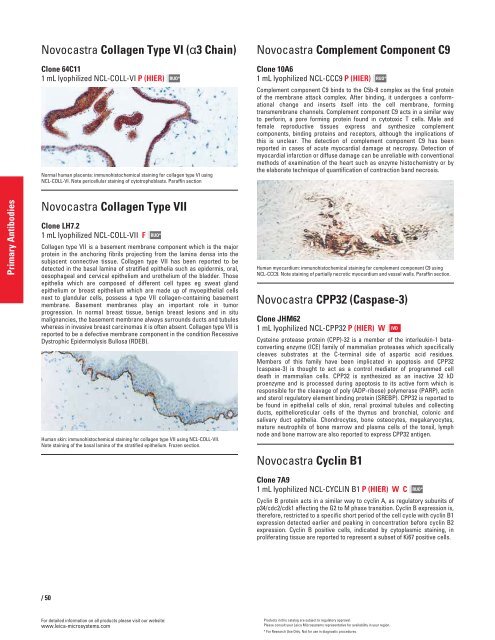
Normal human placenta: immunohistochemical staining for collagen type VI using<br />
NCL-COLL-VI. Note pericellular staining of cytotrophoblasts. Paraffin section<br />
Novocastra Collagen Type VII<br />
Clone LH7.2<br />
1 mL lyophilized NCL-COLL-VII F<br />
/50<br />
RUO*<br />
Collagen type VII is a basement membrane component which is the major<br />
protein in the anchoring fibrils projecting from the lamina densa into the<br />
subjacent connective tissue. Collagen type VII has been reported to be<br />
detected in the basal lamina of stratified epithelia such as epidermis, oral,<br />
oesophageal and cervical epithelium and urothelium of the bladder. Those<br />
epithelia which are composed of different cell types eg sweat gland<br />
epithelium or breast epithelium which are made up of myoepithelial cells<br />
next to glandular cells, possess a type VII collagen-containing basement<br />
membrane. Basement membranes play an important role in tumor<br />
progression. In normal breast tissue, benign breast lesions and in situ<br />
malignancies, the basement membrane always surrounds ducts and tubules<br />
whereas in invasive breast carcinomas it is often absent. Collagen type VII is<br />
reported to be a defective membrane component in the condition Recessive<br />
Dystrophic Epidermolysis Bullosa (RDEB).<br />
Human skin: immunohistochemical staining for collagen type VII using NCL-COLL-VII.<br />
Note staining of the basal lamina of the stratified epithelium. Frozen section.<br />
For detailed information on all products please visit our website:<br />
www.leica-microsystems.com<br />
RUO*<br />
Novocastra Complement Component C9<br />
Clone 10A6<br />
1 mL lyophilized NCL-CCC9 P (HIER)<br />
Complement component C9 binds to the C5b-8 complex as the final protein<br />
of the membrane attack complex. After binding, it undergoes a conformational<br />
change and inserts itself into the cell membrane, forming<br />
transmembrane channels. Complement component C9 acts in a similar way<br />
to perforin, a pore forming protein found in cytotoxic T cells. Male and<br />
female reproductive tissues express and synthesize complement<br />
components, binding proteins and receptors, although the implications of<br />
this is unclear. The detection of complement component C9 has been<br />
reported in cases of acute myocardial damage at necropsy. Detection of<br />
myocardial infarction or diffuse damage can be unreliable with conventional<br />
methods of examination of the heart such as enzyme histochemistry or by<br />
the elaborate technique of quantification of contraction band necrosis.<br />
Human myocardium: immunohistochemical staining for complement component C9 using<br />
NCL-CCC9. Note staining of partially necrotic myocardium and vessel walls. Paraffin section.<br />
Novocastra CPP32 (Caspase-3)<br />
Clone JHM62<br />
1 mL lyophilized NCL-CPP32 P (HIER) W<br />
Cysteine protease protein (CPP)-32 is a member of the interleukin-1 betaconverting<br />
enzyme (ICE) family of mammalian proteases which specifically<br />
cleaves substrates at the C-terminal side of aspartic acid residues.<br />
Members of this family have been implicated in apoptosis and CPP32<br />
(caspase-3) is thought to act as a control mediator of programmed cell<br />
death in mammalian cells. CPP32 is synthesized as an inactive 32 kD<br />
proenzyme and is processed during apoptosis to its active form which is<br />
responsible for the cleavage of poly (ADP-ribose) polymerase (PARP), actin<br />
and sterol regulatory element binding protein (SREBP). CPP32 is reported to<br />
be found in epithelial cells of skin, renal proximal tubules and collecting<br />
ducts, epithelioreticular cells of the thymus and bronchial, colonic and<br />
salivary duct epithelia. Chondrocytes, bone osteocytes, megakaryocytes,<br />
mature neutrophils of bone marrow and plasma cells of the tonsil, lymph<br />
node and bone marrow are also reported to express CPP32 antigen.<br />
Novocastra Cyclin B1<br />
RUO*<br />
Clone 7A9<br />
1 mL lyophilized NCL-CYCLIN B1 P (HIER) W C<br />
Cyclin B protein acts in a similar way to cyclin A, as regulatory subunits of<br />
p34/cdc2/cdk1 affecting the G2 to M phase transition. Cyclin B expression is,<br />
therefore, restricted to a specific short period of the cell cycle with cyclin B1<br />
expression detected earlier and peaking in concentration before cyclin B2<br />
expression. Cyclin B positive cells, indicated by cytoplasmic staining, in<br />
proliferating tissue are reported to represent a subset of Ki67 positive cells.<br />
Products in this catalog are subject to regulatory approval.<br />
Please consult your Leica Microsystems representative for availability in your region.<br />
* For Research Use Only. Not for use in diagnostic procedures.<br />
IVD<br />
RUO*


