Tierärztliche Hochschule Hannover Entwicklung von Methoden zur ...
Tierärztliche Hochschule Hannover Entwicklung von Methoden zur ...
Tierärztliche Hochschule Hannover Entwicklung von Methoden zur ...
Erfolgreiche ePaper selbst erstellen
Machen Sie aus Ihren PDF Publikationen ein blätterbares Flipbook mit unserer einzigartigen Google optimierten e-Paper Software.
SUMMARY<br />
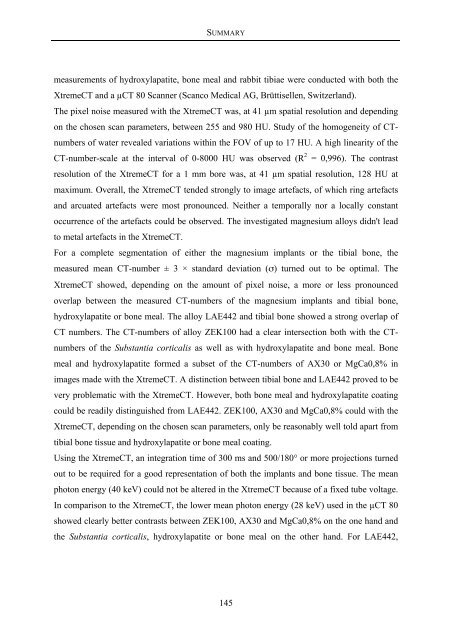
measurements of hydroxylapatite, bone meal and rabbit tibiae were conducted with both the<br />
XtremeCT and a µCT 80 Scanner (Scanco Medical AG, Brüttisellen, Switzerland).<br />
The pixel noise measured with the XtremeCT was, at 41 µm spatial resolution and depending<br />
on the chosen scan parameters, between 255 and 980 HU. Study of the homogeneity of CTnumbers<br />
of water revealed variations within the FOV of up to 17 HU. A high linearity of the<br />
CT-number-scale at the interval of 0-8000 HU was observed (R 2 = 0,996). The contrast<br />
resolution of the XtremeCT for a 1 mm bore was, at 41 µm spatial resolution, 128 HU at<br />
maximum. Overall, the XtremeCT tended strongly to image artefacts, of which ring artefacts<br />
and arcuated artefacts were most pronounced. Neither a temporally nor a locally constant<br />
occurrence of the artefacts could be observed. The investigated magnesium alloys didn't lead<br />
to metal artefacts in the XtremeCT.<br />
For a complete segmentation of either the magnesium implants or the tibial bone, the<br />
measured mean CT-number ± 3 × standard deviation (σ) turned out to be optimal. The<br />
XtremeCT showed, depending on the amount of pixel noise, a more or less pronounced<br />
overlap between the measured CT-numbers of the magnesium implants and tibial bone,<br />
hydroxylapatite or bone meal. The alloy LAE442 and tibial bone showed a strong overlap of<br />
CT numbers. The CT-numbers of alloy ZEK100 had a clear intersection both with the CTnumbers<br />
of the Substantia corticalis as well as with hydroxylapatite and bone meal. Bone<br />
meal and hydroxylapatite formed a subset of the CT-numbers of AX30 or MgCa0,8% in<br />
images made with the XtremeCT. A distinction between tibial bone and LAE442 proved to be<br />
very problematic with the XtremeCT. However, both bone meal and hydroxylapatite coating<br />
could be readily distinguished from LAE442. ZEK100, AX30 and MgCa0,8% could with the<br />
XtremeCT, depending on the chosen scan parameters, only be reasonably well told apart from<br />
tibial bone tissue and hydroxylapatite or bone meal coating.<br />
Using the XtremeCT, an integration time of 300 ms and 500/180° or more projections turned<br />
out to be required for a good representation of both the implants and bone tissue. The mean<br />
photon energy (40 keV) could not be altered in the XtremeCT because of a fixed tube voltage.<br />
In comparison to the XtremeCT, the lower mean photon energy (28 keV) used in the µCT 80<br />
showed clearly better contrasts between ZEK100, AX30 and MgCa0,8% on the one hand and<br />
the Substantia corticalis, hydroxylapatite or bone meal on the other hand. For LAE442,<br />
145







![Tmnsudation.] - TiHo Bibliothek elib](https://img.yumpu.com/23369022/1/174x260/tmnsudation-tiho-bibliothek-elib.jpg?quality=85)
