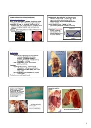
Oral and Poster Abstracts
Oral and Poster Abstracts
Oral and Poster Abstracts

Create successful ePaper yourself
Turn your PDF publications into a flip-book with our unique Google optimized e-Paper software.
was to evaluate, by serology, prevalence of Coxiella burnetii infection<br />
in herds of Caltanissetta district. 124 of 289 herds were selected, with<br />
stratified r<strong>and</strong>om sampling, distributed in four parts of Caltanissetta<br />
district. Particularly 71 herds were chosen from Caltanissetta territory,<br />
9 from Gela, 29 from Mussomeli <strong>and</strong> 15 from San Cataldo. 1711 sera<br />
were collected to test the presence of specific antibodies to C. burnetii.<br />
Sera were tested by a commercial available ELISA (Chekit Q-fever;<br />
IDEXX Laboratoires). Results showed serological positivity in 60/124<br />
(48,4%) herds with 118 positive sera/1711 (6,9%).<br />
727 Apparent Prevalence of Antibodies to Coxiella burnetii (Q<br />
Fever) in Bulk Tank Milk of Dairy Herds in the Walloon<br />
Region of Belgium<br />
G. Czaplicki 1 , J. Houtain 1 , C. Manteca 2 , C. Saegerman 3<br />
1<br />
ARSIA, Diagnostic Center for Animal Diseases, Loncin, Belgium<br />
2<br />
CEVA santé Animale, Diagnostic Center for Animal Diseases,<br />
Libourne, France<br />
3<br />
University of Liege - Faculty of Veterinary Medicine, Epidemiology<br />
<strong>and</strong> Risk Analysis applied to Veterinary sciences, Faculty of, Liege,<br />
Belgium<br />
Objectives: Q Fever is a zoonotic disease caused by Coxiella burnetii,<br />
an obligate intracellular bacteria. Ruminants (sheep, goats <strong>and</strong> cattle)<br />
remain the major natural reservoir for human infection. The aim of the<br />
present study was on the one h<strong>and</strong>, to evaluate the apparent prevalence<br />
in the Walloon dairy herds <strong>and</strong> on the other h<strong>and</strong>, to identify herd level<br />
risk factors <strong>and</strong> clinical signs statistically associated with seropositivity<br />
in the population of r<strong>and</strong>omly selected Walloon dairy cattle.<br />
Material <strong>and</strong> methods: 566 herds were r<strong>and</strong>omly selected according to a<br />
classical methodology (Jenicek <strong>and</strong> Cléroux, 1987). Herd level predictors<br />
data were collected from a questionnaire on farm demographics,<br />
management practices <strong>and</strong> observed clinical signs during the twelve last<br />
months. Two hundred <strong>and</strong> six farms responded on a voluntary basis to this<br />
questionnaire <strong>and</strong> submitted a sample of bulk tank milk sampled in<br />
February 2006. Bulk milks were centrifuged <strong>and</strong> stored at 20°C until<br />
testing for antibodies to Coxiella burnetii with a commercial Indirect Elisa<br />
test (LSI). Apparent prevalence was estimated with a ninety-five percent<br />
confidence intervals (95% CI) assuming a binomial exact distribution.<br />
Statistical analysis of the data was done using a chi square test <strong>and</strong> the<br />
tendency of each parameter to become a risk factor was evaluated by an<br />
odds ratio calculation with 95% confidence intervals (logarithmic<br />
approximation); a P value < 0.05 was considered significant.<br />
Results: Global apparent prevalence: 54.9 % (95% CI : 47.8-61.8%) of<br />
herds show serological evidence of Coxiella burnetii infection. Clinical<br />
signs as abortion in heifers <strong>and</strong> in cows, stillborn or weak calves are<br />
statistically associated with seropositivity. Risk factors associated with<br />
seropositivity are free stalling <strong>and</strong> watering with well water while<br />
blocked stalling, watering with tap water <strong>and</strong> disinfection of sheds act<br />
as protectors of infection.<br />
Conclusions: This first report on Q Fever infection prevalence in<br />
Walloon dairy herds indicates a quite high level of infection. Seropositivity<br />
at herd level does not mean shedding of Coxiella burnetii, but<br />
identifies herds where at least 10% of lactating animals are sero-positive<br />
to C. burnetii. A significant association exists between sero-positivity<br />
<strong>and</strong> typical clinical signs of Q Fever observed in those herds. Some risk<br />
factors <strong>and</strong> some protectors are identified on a statistical basis.<br />
Key words: bovine, Q Fever, serology, prevalence<br />
728 Identification of Bovine Viral Diarrhoea Virus<br />
Mimotopes/Epitopes through Selection from a Peptide 12-mer<br />
Phage Display Library<br />
A. Zamit 1 , M. Ostrowski 2 , N. Fondevila 1 , O. Lopez 3 , A. Bratanich 4 ,<br />
S. Romera 1<br />
1<br />
Instituto Nacional de Tecnología Agropecuaria-INTA, BUENOS<br />
AIRES, Argentina<br />
2<br />
Institut Curie, Paris, France<br />
3<br />
Northern Michigan University, Marquette, United States<br />
4<br />
Facultad de Cs Veterinarias-UBA, BUENOS AIRES, Argentina<br />
Bovine Viral Diarrhoea Virus (BVDV) is a worldwide distributed<br />
pestivirus that causes important economical losses to cattle industry.<br />
Argentine is no exception with a population of 50 million bovines <strong>and</strong><br />
high seroprevalences up to 90%. The aim of our study is epitope<br />
mapping of immunodominant E2 protein of BVDV using a peptide 12mer<br />
phage display library.<br />
Materials <strong>and</strong> Methods: A peptide 12-mer phage display commercial<br />
library was depleted of mimotopes resembling BVDV proteins except<br />
those corresponding to E2, by using serum from a calf immunized<br />
against recombinant BVDV-E2 BDV. Then, a positive selection was<br />
performed with affinity purified antibodies against BVDV strain NADL.<br />
Individual phage clones were amplified <strong>and</strong> displayed peptide sequences<br />
analyzed using CLUSTALX with Tudos matrix. Four peptides were<br />
designed for immunization purposes <strong>and</strong> sent to synthesize. Two bovines<br />
<strong>and</strong> 4 groups of guinea pigs were vaccinated with peptides coupled to<br />
KLH. Also, phages displaying chosen peptides were amplified <strong>and</strong> used<br />
to immunize mice. Sera were analyzed by peptide-ELISA, phage-<br />
ELISA, indirect ELISA <strong>and</strong> seroneutralization.<br />
Results: Enrichment of specific clones was achieved in 4 rounds of panning<br />
<strong>and</strong> 30 clones were individually amplified. Multiple sequence alignment<br />
analysis with modified parameters <strong>and</strong> substitution matrix allowed a better<br />
clustering of peptides. Considering clusters, peptide frequency <strong>and</strong><br />
hydrophobicity <strong>and</strong> aminoacids representativity in library, 4 rationally<br />
designed peptides with immunogenic properties were obtained. Satisfactory<br />
antibody responses against peptides were observed in mice immunized with<br />
phages, <strong>and</strong> bovines <strong>and</strong> guinea pigs vaccinated with synthetic peptides.<br />
Also, antibodies produced in mice recognized synthetic peptides. Sera from<br />
immunized animals were either citotoxic or not neutralizing. Indirect<br />
ELISA showed high background <strong>and</strong> was not conclusive.<br />
Conclusions: It was possible to obtain immunogenic peptides<br />
resembling epitopes of BVDV using a peptide 12-mer phage display<br />
library as mapping strategy. Epitope mapping is imperative for<br />
improving local vaccines design <strong>and</strong> control.<br />
Key words: BVDV, mimotope, phage display, E2<br />
729 Amplification of Inv Gene of Salmonalla Serotypes by<br />
Polymerase Chain Reaction (PCR) as a Specific Method of<br />
Detection of Salmonellae<br />
T. Zahraei Salehi 1 , M. Mahzonieh 2 , A. Ashrafi 1<br />
1<br />
Faculty of Veterinary Medicine, University of Tehran, Microbiology,<br />
Tehran, Iran<br />
2<br />
Faculty of Veterinary, Pathobiology, Shahrekord, Iran<br />
Background: Detection of inv gene in Salmonella serotypes by PCR.<br />
Materials <strong>and</strong> Methods: Sixty Salmonella strains were isolated from<br />
animals <strong>and</strong> human sources. In this research, at first 60 isolated<br />
Salmonella from animals <strong>and</strong> human were tested by biochemical tests<br />
such as carbohydrate utilization tests <strong>and</strong> urease test <strong>and</strong> then were<br />
serogrouped by Salmonella O anisera. DNA of isolated Salmonella<br />
were extracted by Holmes <strong>and</strong> Quigley method. Two primers (St139<br />
<strong>and</strong> St141) <strong>and</strong> PCR reagents from fermantas company were used for<br />
amplication of inv gene. PCR reaction was carried out in Master cycle<br />
(Eppendorf). The PCR product were loaded into 1.2% agarose gel <strong>and</strong><br />
electrophoresed for 60 minutes at 120 V.<br />
Results: All isolates showed biochemical properties of Salmonellae. In<br />
PCR assay, target gene (invA gene) with 284 bp size were observed in all<br />
the strains, which is corresponded with size of b<strong>and</strong> of positive control <strong>and</strong><br />
DNA marker. So, all of the strains in this survey had invA gene.<br />
Conclution: According to the results of this study PCR method based<br />
on inv gene is useful to rapid identification of Salmonella serotypes.<br />
Key words: Salmonella, invA gene, PCR<br />
730 Toxoplasmosis in a Commercial Dairy Farm in Turkey<br />
S. Gazyagci 1 , C. Babur 2 , S. Kilic 2 , B. Celebi 2 , A. Gazyagci 2 ,<br />
B. Yagci 1<br />
1<br />
Faculty of Veterinary in Kirikkale University, Department of<br />
Internal Medicine, Kirikkale, Turkey<br />
2<br />
National Research Center, Epidemiology <strong>and</strong> Public Health,<br />
Ankara, Turkey<br />
Blood samples were taken from 742 dairy cattles in a commercial farm.<br />
Antibody titers were determined against Toxoplasma gondii by Sabine-<br />
Feldman Dye method. Serological examination revealed 640 cattle to<br />
be seropositive. Among these sera the numbers of seropositive samples<br />
at certain dilution steps were as follows: 268 at 1/16, 184 at 1/64, 112<br />
at 1/256 <strong>and</strong> 76 at 1/1024. Three cats could circulate in the warehouse<br />
in the farm. All sera were tested with Sabin-Feldman Dye test for<br />
Toxoplasma gondii spesific antibodies. Antibody titers were 1/256 all<br />
of them.This study investigates the seroprevalence of Toxoplasma<br />
gondii in dairy cattle from a commercial farm in Turkey.<br />
Key words: cattle, Sabin Feldman dye test, Toxoplasma gondii<br />
Infectious <strong>and</strong> Zoonotic Deseases (Public Health) 109