Oral and Poster Abstracts
Oral and Poster Abstracts
Oral and Poster Abstracts

Create successful ePaper yourself
Turn your PDF publications into a flip-book with our unique Google optimized e-Paper software.
position. A fusiform skin incision was created around the protruding<br />
mass <strong>and</strong> the hernial ring <strong>and</strong> abdominal wall were exposed. The<br />
hernial ring was excised <strong>and</strong> the protruding mass was identified as<br />
the abomasum. A large part of the abomasal body (including the<br />
curvatura major <strong>and</strong> minor) was involved in the hernia. After<br />
removal of all necrotic tissue, the abomasum was reconstructed<br />
using an end-to-end anastomosis. The surgical procedure was<br />
accompanied with a severe haemorrhage inducing a decrease of the<br />
packed cell volume from 24 to 17%. Recovery from anaesthesia was<br />
uneventful. An intensive intravenous supportive therapy was<br />
installed afterwards (lactated ringer solution supplemented with<br />
glucose, calcium <strong>and</strong> potassium chloride). Apart from some oedema<br />
around the abdominal wound, the cow had fully recovered after 10<br />
days <strong>and</strong> was dismissed from the clinic. The subsequent lactation<br />
was uneventful.<br />
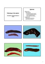
Abomasal fistulation has rarely been described as a complication of<br />
omentopexy. This fistulation might be caused by inadvertent<br />
penetration of the abomasal lumen during a st<strong>and</strong>ard omentopexy<br />
procedure. Eventually, abomasal ulcerations can be induced by the<br />
chronic inflammation following the omentopexy, creating a<br />
fistulation through the abdominal wall. The combination of an<br />
abomasal fistula <strong>and</strong> an abdominal hernia can end in a complete<br />
herniation of the abomasum, requiring an end-to-end anastomosis.<br />
424 Laparoscopy-assisted Biopsy of Mesenteric Lymph Nodes in<br />
Calves<br />
T. Seeger1 , P. Berisha1 , K. Koehler2 , M. Buelte3 , K. Doll1 1 University of Giessen, Clinic of Ruminants, Giessen, Germany<br />
2 University of Giessen, Institute for Veterinary Pathology, Giessen,<br />
Germany<br />
3 University of Giessen, Institute of Veterinary Food Science, Giessen,<br />
Germany<br />
Objectives of study: Examination of mesenteric lymph nodes is still<br />
the “gold-st<strong>and</strong>ard” for diagnosis of infections with Mycobcterium<br />
avium ssp. paratuberculosis in cattle. Results of other studies have<br />
shown that this bacterium is also detectable in mesenteric lymph nodes<br />
of young animals. Aim of this study was to validate a laparoscopic<br />
method for the biopsy of these lymph nodes in calves.<br />
Material <strong>and</strong> Methods: The study was performed on 15 Holstein<br />
calves. All calves were between 2 <strong>and</strong> 4 weeks of age. During the 3<br />
week study period the animals were housed in the clinic. To identify<br />
complications after laparoscopic surgery clinical examinations were<br />
performed twice daily <strong>and</strong> blood samples were taken on several<br />
study days. At the end of the study period all calves were euthanised<br />
<strong>and</strong> a necropsy was performed at the Institute for Veterinary<br />
Pathology. The laparoscopy-assisted biopsy was performed on day 7<br />
of the study. Before surgery all calves were treated with antibiotics<br />
<strong>and</strong> antiphlogistics for one time. Detomidine (0.05 mg/kg im) <strong>and</strong><br />
ketamine (6 mg/kg iv) were admistered for anesthesia. The calves<br />
were fixed on an operating table in right lateral recumbency. The left<br />
abdominal wall was prepared for aseptic surgery. Three stab<br />
incisions of the skin were performed <strong>and</strong> a Verres needle was<br />
inserted into the abdomen to create a pneumoperitoneum. Three<br />
magnetic valve trokars were inserted such that a rigid laparoscope<br />
<strong>and</strong> 2 forceps could be introduced into the abdominal cavity. After<br />
visualisation <strong>and</strong> fixation of the cecum the ileocecal lymph node was<br />
fixated <strong>and</strong> a biopsy was taken with a special spoon forceps. Same<br />
procedure was performed for biopsy of one lymph node adjacent to<br />
the caudal part of the jejunum. At the end of surgery the 3 skin<br />
incisions were closed by a simple suture.<br />
Results: No complications due to the anesthesia or the<br />
pneumoperitoneum were observed. The average operation time was<br />
26.2 minutes with a range from 18 to 37 minutes. The biopsy of a<br />
jejunal lymph node was successful in all calves. In one calf only the<br />
biopsy of the ileocecal lymph node was impossible. No major<br />
postoperative complications were observed. During the necropsies no<br />
signs of peritonitis or wound complications were detected.<br />
Conclusions: The results of this study show that the described<br />
laparoscopic method for biopsy of mesenteric lymph nodes in calves is<br />
well suited to obtain these important specimens for paratuberculosis<br />
diagnosis.<br />
Key words: laparoscopy, lymph node biopsy, paratuberculosis, calf<br />
425 Two-dimensional <strong>and</strong> M-mode Echocardiographic<br />
Parameters Measured in Fifteen Healthy Calves<br />
258 XXV. Jubilee World Buiatrics Congress 2008<br />
N. Cesbron 1 , V. Charot 2 , A. Dorizon 2<br />
1 National Veterinary School of Nantes, Unité de Médecine des<br />
Animaux d’Elevage, Nantes, France<br />
2 National Veterinary School of Nantes, Unité d’Imagerie Médicale,<br />
nantes, France<br />
Objectives of study: Within ultrasonography procedures,<br />
echocardiography has not been studied as extensively in cattle as in<br />
pets. Among all the studies dealing with echocardiography in cattle,<br />
most of them were mainly focused either (i) on examinations of cardiac<br />
affections (like endocarditis, pericarditis or congenital abnormalities)<br />
or (ii) in healthy adult cattle. Few studies have been made in calves.<br />
Therefore, the aim of this study was to obtain reference values for the<br />
echocardiographic parameters in healthy calves using both twodimensional<br />
real-time <strong>and</strong> M-Mode echography.<br />
Materials <strong>and</strong> methods: To reach this goal, fifteen Holstein<br />
clinically healthy calves were used. Their ages ranged from 2 weeks<br />
to 1 month. The examinations were made while the calves were<br />
st<strong>and</strong>ing using a 2.5 MHz convex transducer. The heart was<br />
examined ultrasonographically on the right <strong>and</strong> then the left side<br />
with the transducer in four positions: caudal long, caudal short <strong>and</strong><br />
cranial long axes on the right side; caudal long axe on the left side.<br />
The variables were measured in a short axis view of the heart <strong>and</strong><br />
both two-dimensional (2D) <strong>and</strong> cursor- directed time-motion (M)<br />
modes were used. Measurements, at systole (s) <strong>and</strong> diastole (d), of<br />
the left ventricular internal diameter (respectively LVDs <strong>and</strong> LVDd),<br />
<strong>and</strong> of the interventricular septum thickness (respectively IVSs <strong>and</strong><br />
IVSd) were obtained according to the previous described procedure.<br />
The left atrial diameter (LAD), the aortic root diameter (AOD) <strong>and</strong><br />
their ratio [LAD/AO] were described.<br />
Results: For the ventricular internal diameter at systole <strong>and</strong> diastole,<br />
the following results (in mm) were obtained with respectively [LVDd:<br />
44.57 ± 6.53], [LVDs: 30.07 ± 5.82]. For the interventricular septum<br />
thickness, we observed at systole <strong>and</strong> diastole (in mm) [LVSd: 11.90 ±<br />
2.33], [IVSs: 16.64 ± 2.19]. Lastly, concerning the left atrial <strong>and</strong> the<br />
aortic diameters, we observed (in mm) [AOD: 31.18 ± 3.02], [LAD:<br />
34.31 ± 4.26] leading to a ratio [LAD/AO: 1.11 ± 0.17].These results<br />
are slightly lower than those reported by a previous study performed by<br />
Amory et al. on older calves (> 2 months).<br />
Conclusion: The echocardiographic reference values obtained on these<br />
healthy calves aged from less than one month were similar to those<br />
classically reported for dogs of similar body weight (large breed size).<br />
Key words: echocardiography, calves, M-mode <strong>and</strong> two-dimensional<br />
echocardiography<br />
426 Measurements of Dimensions of Holstein-Friesian Calves<br />
Using Computed Tomography in order to Compare Different<br />
Obstetrical Traction Methods<br />
C. Heun 1 , G. Tsousis 1 , M. Becker 1 , J. Rieder 2 , H. Bollwein 1<br />
1 University of Veterinary Medicine Hannover, Clinic for Cattle,<br />
Hannover, Germany<br />
2 University of Veterinary Medicine Hannover, Small Animal Clinic,<br />
Hannover, Germany<br />
The aim of this study was to measure the difference in calves’<br />
dimensions resulting from different obstetrical methods. 20 stillborn<br />
calves (41.1 +/- 3.7 kg) were measured objectively by computed<br />
tomography. The calves were placed stretched on their right side<br />
simulating the simultaneous traction with both front legs on the same<br />
level (0D), the alternate traction with 5 cm difference between the level<br />
of the legs (5D) <strong>and</strong> the alternate traction with 10 cm difference<br />
between the level of the legs (10D). All measurements were carried out<br />
with special software. For the statistical analysis repeated measures<br />
ANOVA was used. For each traction the maximum cross-sectional area<br />
on the level of the forehead (ma-F) <strong>and</strong> on the elbows, the biggest width<br />
of the shoulders on the level of the tuberculum majus (width-tm) <strong>and</strong><br />
the perimeter on the level of the forehead (peri-F) were measured. The<br />
ma-F reduced from traction 0D to 5D with a mean difference of 2.9 ±<br />
4.1 cm (p=0.005), from 5D to 10D with 8.8 ± 5.8 cm (p