Oral and Poster Abstracts
Oral and Poster Abstracts
Oral and Poster Abstracts

You also want an ePaper? Increase the reach of your titles
YUMPU automatically turns print PDFs into web optimized ePapers that Google loves.
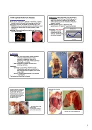
499 Secondary Hepatogenous Photosensitization in Buffalos due<br />
to the Excessive Accumulation of Hepatic Copper<br />
A. Minervino 1 , R. Barreto júnior 2 , F. Rodrigues 1 , L. Reis 1 ,<br />
R. Ferreira 1 , S. Headley 3 , E. Ortolani 1<br />
1<br />
College of Veterinary Medicine, University of Sao Paulo,<br />
Department of Clinical Sciences, Sao Paulo, Brazil<br />
2<br />
Rural Federal University of Semi-Arid, Mossoró, Brazil<br />
3<br />
Faculty of Veterinary Medicine, University of Helsinki, Helsinki,<br />
Finl<strong>and</strong><br />
Four 1.5 yr-old, male, murrah buffaloes, maintained during six months<br />
with high Cu intake <strong>and</strong> without direct solar exposure, were transported<br />
to regular farming conditions. Shortly after sunlight exposure, all<br />
buffalos demonstrated clinical manifestations characteristic of<br />
photosensitization: anorexia, apathy <strong>and</strong> severe cutaneous lesions.<br />
Blood samples were obtained before solar exposure (T0), during the<br />
clinical manifestations (T1), <strong>and</strong> after recovery (T2) to evaluate<br />
haematological (PCV, Hb, RBC <strong>and</strong> WBC) <strong>and</strong> biochemical (total<br />
protein, albumin, g-glutamil transferase (gGT), aspartate amino<br />
transferase (AST), creatine kinase (CK), urea, <strong>and</strong> creatinine)<br />
parameters. Skin samples obtained by incision biopsy at T0 <strong>and</strong> T1<br />
were processed for routine histopathology. Treatment consisted of<br />
removal from sunlight exposure <strong>and</strong> aqueous Zinc oxide solution.<br />
Three animals recovered fullly <strong>and</strong> 1 died. The hepatic Cu<br />
concentrations <strong>and</strong> gGT <strong>and</strong> AST activities before solar exposure were<br />
above normal values. For haematological parameters, only RBC<br />
demonstrated significant difference, being reduced at T1 <strong>and</strong> returning<br />
to normality at T2. A similar pattern was observed with the total protein<br />
<strong>and</strong> albumin; but an opposite trend occurred with CK activity. The<br />
buffalo that succumbed had the highest liver of Cu concentration <strong>and</strong><br />
more extensive macroscopic skin lesions. At T0 the buffalos had<br />
generalized alopecia, but insignificant histological alterations<br />
(acantholysis <strong>and</strong> neovascularization). However, at T1<br />
histopathological examination revealed hepatic photosensitization<br />
characterized by diffused, moderate parakeratotic hyperkeratosis,<br />
multifocal moderate acantholysis, degeneration <strong>and</strong> necrosis of<br />
squamous epithelial cells, sweat gl<strong>and</strong> atrophy, sebaceous gl<strong>and</strong><br />
hyperplasia, <strong>and</strong> dermal edema. Although the buffalos demonstrated a<br />
liver insufficiency at T0, the skin lesions indicative of secondary<br />
hepatogenous photosensitization were only triggered after the solar<br />
exposure. In this case, a high Cu intake induced hepatic lesions that<br />
may have caused inefficient metabolization of phylloerythrin resulting<br />
in secondary photosensitization after the sunlight exposure.<br />
Research supported by FAPESP<br />
Key words: buffaloes, hyperkeratosis, photosensitization, hepatic<br />
lesions<br />
500 Trace Elements in Cattle: Copper <strong>and</strong> it`s “Friends”<br />
A. Mueller, B. Freude, M. Scholz, M. Weiss<br />
IDEXX, Vet Med Labor GmbH, Ludwigsburg, Germany<br />
Prophylactic procedures to prevent diseases become more <strong>and</strong> more<br />
important in cattle rearing. Adequate supply in trace elements is<br />
required. Copper (Cu) deficiency is regarded as the second most<br />
common mineral deficiency of cattle in the World (Telfer et al, 1996;<br />
Black und French, 2004). Two different copper deficiencies are known:<br />
a primary copper deficiency induced by a simple deficiency of copper<br />
in the diet, <strong>and</strong> a secondary, resulting from the reduction in copper<br />
absorption or utilization by the antagonistic effects of molybdenum<br />
(Mo) <strong>and</strong> iron (Fe). 340 cattle samples were analysed as serum/plasma<br />
pairs regarding the concentration of Cu, Mo <strong>and</strong> Fe with ICP-AES<br />
(Inductively Coupled Plasma Atomic Emission Spectroscopy) during<br />
July 2006 until March 2007. The Cu-plasma analyses showed that only<br />
24% of these samples were below the reference value for Cu (80<br />
Ïg/dL). Compared to these results, 84% of the corresponding serum<br />
samples showed a Cu deficiency (below 80 µg/dL). Serum clotting<br />
reduces Cu by up to 80% compared to the plasma values (in<br />
confirmation to Laven & Livesey, 2006). In contrast to copper the<br />
analyses of Mo in bovine plasma <strong>and</strong> serum resulted in comparable<br />
values. 4500 bovine serum <strong>and</strong> plasma samples were analysed for Mo.<br />
80% of Mo-concentrations were below 26 µg/L. Only a few samples (<<br />
1%) showed higher values (max. 3300 µg/L). A correlation between<br />
low Cu <strong>and</strong> high Mo values in plasma, or low Cu values with high Fe<br />
concentrations could not be found. Additionally, hair analyses of Cu (N<br />
= 173) <strong>and</strong> Mo (N = 99) were performed. 85 % of the hair samples (3-<br />
30 XXV. Jubilee World Buiatrics Congress 2008<br />
times analyses of each hair sample) showed Cu values between 5 <strong>and</strong><br />
20 mg/kg (49%: 6,6-10,4 mg/kg; 4 %: below 6,6 mg/kg), 15 % of the<br />
samples showed higher values up to 100 mg/kg). The Mo<br />
concentrations in the hair samples varied between 0 up to 800 µg/kg or<br />
even higher). The comparison between Cu <strong>and</strong> Mo in hair (N = 99), or<br />
Cu hair/plasma (N = 107) <strong>and</strong> Mo hair/plasma (N = 70) showed no<br />
correlation. The analysed cattle samples with low copper plasma<br />
concentrations showed mainly a primary copper deficiency. A<br />
secondary copper deficiency caused by a molybdenum intoxication<br />
could not be found in this set of samples.<br />
501 Effect of chronic glucose infusion on lactation performance<br />
<strong>and</strong> metabolic profiles in dairy cows<br />
B. Al-Trad 1 , K. Reisberg 1 , T. Wittek 2 , A. Alkaassem 2 , G. Gäbel 1 ,<br />
M. Fürll 2 , J.R. Aschenbach 1<br />
1 Faculty of veterinary medicine, University of Leipzig, Institute of<br />
Animal Physiology, Leipzig, Germany<br />
2 Faculty of veterinary medicine, University of Leipzig, Medizinische<br />
Tierklinik, Leipzig, Germany<br />
Objective: To characterize the metabolic adaptations that occur during<br />
gradual increases of glucose supply via prolonged i.v. glucose<br />
infusions in dairy cows.<br />
Material & methods: Twelve midlactation dairy cows were assigned<br />
r<strong>and</strong>omly to continuous i.v. infusions of either saline (control group,<br />
n=6) or 40% glucose solutions (treatment group, n=6) for 28 d. The<br />
infusion dose started at 1.25% of the daily energy (NEL) requirement<br />
<strong>and</strong> then gradually increased until a maximum dose of 30% NEL<br />
requirement was achieved at d 23. Then infusion dose was maintained<br />
at 30% for 5 d. The treatment (feed plus infusion) was hypercaloric but<br />
isonitrogenous. Liver <strong>and</strong> skeletal muscle biopsies were taken on d 0, 8,<br />
16, 24, <strong>and</strong> 32. Blood samples were taken every 2 d at 10:00 a.m. <strong>and</strong><br />
additional 24-h blood samples (6-h intervals) were taken before each<br />
biopsy. Milk <strong>and</strong> urine samples were also taken on the biopsies days.<br />
Results: No changes occurred in daily feed intake <strong>and</strong> milk production,<br />
milk lactose <strong>and</strong> urea concentrations. Milk protein percentage <strong>and</strong> yield<br />
tended to increase during the high infusion dose (i.e. 20 & 30% NEL<br />
requirement). Decrease in milk fat percentage <strong>and</strong> yields were<br />
observed in both groups; however, the decrease was numerically higher<br />
in the treatment cows. Body weight <strong>and</strong> BFT increased in the treated<br />
group. Small <strong>and</strong> occasional increases in blood glucose <strong>and</strong> insulin<br />
concentrations were observed in the blood samples taken at 10:00 a.m.<br />
every 2 d. However, at infusion of 30% NEL requirement, five out of<br />
six treated cows had blood glucose concentrations >8 mM at 16:00<br />
p.m., i.e. 1 h postpr<strong>and</strong>ially. Glucose infusion decreased serum NEFA,<br />
BHBA <strong>and</strong> BUN concentrations. Serum liver enzymes, cholesterol,<br />
bilirubin, K <strong>and</strong> Ca concentrations were not affected by infusion. Less<br />
than 2% changes were observed in serum P, Cl <strong>and</strong> Na concentrations.<br />
Glucoseuria was detected during the maximum infusion dose. Liver<br />
glycogen increased gradually to reach plateau on d 16 before it fell<br />
back to baseline values on d 32. Skeletal muscle glycogen increased<br />
<strong>and</strong> liver total lipid tended to increase during the high dosage of<br />
glucose infusion.<br />
Conclusions: Dairy cows on an energy-balanced diet do not direct<br />
excess glucose to increased lactation performance. Excess glucose is<br />
transiently stored as glycogen in the liver <strong>and</strong>, predominantly,<br />
transferred to body fat. At high dosages, glucose is also stored in<br />
skeletal muscle as glycogen <strong>and</strong> excreted via the urine.<br />
502 Biochemical Profile of Cattle with Induced Hypocalcaemia<br />
<strong>and</strong> Subsequently Treated with an Enriched Calcium<br />
Solution<br />
R. Barreto júnior 1 , A. Minervino 2 , F. Rodrigues 2 , E. Meira júnior 2 ,<br />
R. Ferreira 2 , A. Lima 3 , C. Mori 2 , E. Ortolani 2<br />
1 Rural Federal University of Semi-Arid, Mossoró, Brazil<br />
2 College of Veterinary Medicine, University of Sao Paulo,<br />
Department of Clinical Science, Sao Paulo, Brazil<br />
3 Vallée S.A., Sao Paulo, Brazil<br />
Twelve two years-old Holstein heifers were used to study the<br />
biochemical profile during the induction <strong>and</strong> recovery of<br />
hypocalcaemia. The picture was induced by continuous infusion of a<br />
EDTA solution (5% <strong>and</strong> pH 7.4) into the jugular vein at the speed of<br />
220 mL/h until the animals presented definitive clinical signs of<br />
hypocalcaemia such as sternal or lateral recumbency; then the infusion