Oral and Poster Abstracts
Oral and Poster Abstracts
Oral and Poster Abstracts

Create successful ePaper yourself
Turn your PDF publications into a flip-book with our unique Google optimized e-Paper software.
731 Detection <strong>and</strong> Investigation on the within-farm Transmission<br />
of Bovine Herpesvirus-1 in Farms Located in the Trace<br />
District of Turkey<br />
A. Unver 1 , N. Turan 2 , H. Yilmaz 2<br />
1 Farmamag, Firuzkoy Yolu, Baglar Sokak, Avcilar, Istanbul, Turkey<br />
2 University of Istanbul, Veterinary Faculty, Department of Virology,<br />
Avcilar, Istanbul, Turkey<br />
The aim of this study was to detect BHV-1 infection <strong>and</strong> to investigate<br />
within-farm transmission in farms located in the Trace district of<br />
Turkey. For this, 3 dairy farms having the animals over 20 were<br />
selected from the Marmara region. Two visits were made to these farms<br />
6-7 months intervals. At each visit, the nasal swabs <strong>and</strong> blood from<br />
each animal <strong>and</strong> the milk sample from milking cows were taken. Blood<br />
sera were analysed by ELISA (IDEXX, gB blocking) for the presence<br />
of antibodies to BHV-1. Whereas the PCR was used to detect BHV-1<br />
DNA in the nasal swabs <strong>and</strong> milk samples. In samples taken during the<br />
first visit, antibodies to BHV-1 were detected in 13 of 89 animals in 3<br />
farms. 4 of 27 (14.8%) animals, 6 of 21 (28.7%) animals <strong>and</strong> 3 of 41<br />
(7.3%) animals were found to be seropositive in farms-1, 2 <strong>and</strong> 3,<br />
respectively. BHV-1 DNA was detected in nasal swabs of 2 animals<br />
<strong>and</strong> milk sample of one animal in farm-1, milk sample of 1 animal in<br />
farm-2 <strong>and</strong> nasal swabs of 6 animals in farm-3. In samples taken during<br />
the second visit, antibodies to BHV-1 were detected in 18 of 71 animals<br />
in 3 farms. 6 of 24 (25%) animals, 6 of 17 (35.2%) animals <strong>and</strong> 6 of 30<br />
(20%) animals were found to be seropositive in farms-1, 2 <strong>and</strong> 3,<br />
respectively. BHV-1 DNA was detected in nasal swabs of 3 animals in<br />
farm-1, nasal swabs of 2 animals <strong>and</strong> milk sample of 1 animal in farm-<br />
2. No BHV-1 DNA was detected in the samples taken from the farm-3.<br />
In conclusion, the BHV-1 infection was detected by ELISA <strong>and</strong> PCR in<br />
all 3 farms. Some of the animals found to be seronegative by ELISA<br />
were positive on PCR <strong>and</strong> these animals might be either seronegative<br />
carriers or acutely infected. Therefore, it would be better to check the<br />
animals in the farms by both test ELISA <strong>and</strong> PCR. Also care must be<br />
taken to test new animals <strong>and</strong> separating the youngs from the adults.<br />
Key words: bovine, herpesvirus-1, transmission, PCR, ELISA, Turkey<br />
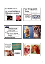
733 Outbreak of Foot-<strong>and</strong>-Mouth Disease with High Fatality Rate<br />
in Young Lambs in Tabriz - Iran<br />
G. Noursaadat 1 , M. Tooloei 2 , F. Rezazadeh 2<br />
1 Large Animal Clinician, Tabriz, Iran<br />
2 University of Tabriz, Faculty of Veterinary Medicine, Department of<br />
Clinical Science, Tabriz, Iran<br />
Foot-<strong>and</strong>-mouth disease (FMD) is caused by an aphthovirus (family<br />
picornaviridae) <strong>and</strong> affects all cloven foot animals. It is endemic in<br />
Africa, Asia, South America <strong>and</strong> parts of Europe. The disease in adult<br />
sheep usually causes milder clinical signs than in cattle or pigs, <strong>and</strong> is<br />
often subtle enough to go undiagnosed. In contrast, FMD in lambs has<br />
been reported to cause high mortality during field outbreaks. From<br />
January to April 2007, five to fifty days-old lambs of the approximately<br />
all of the flocks pertaining to the east region of Tabriz affected with a<br />
highly fatal disease. The morbidity <strong>and</strong> mortality rates in lambs of each<br />
flock in this outbreak were about 10-50 <strong>and</strong> 70-80%, respectively.<br />
Adult sheep had also been affected, but the disease in sheep was very<br />
mild <strong>and</strong> with less frequent <strong>and</strong> extent than lambs. All affected lambs<br />
were clinically examined. Observed clinical signs include: severe<br />
tachycardia, high fever (up to 42˚C), tachypnea, dispnea, salivation <strong>and</strong><br />
drooling, development of vesicular lesions in interdigital <strong>and</strong> coronary<br />
region of all four foot <strong>and</strong> in mouth especially over the tongue <strong>and</strong> lips<br />
as well as lameness. Most of the affected sheep were also suffering<br />
from lameness. At the necropsy of at least one hundred died lambs,<br />
hyperemia <strong>and</strong> hepatization of lungs especially in the apical <strong>and</strong> ventral<br />
lobes, swelling <strong>and</strong> enlargement of mediastinal lymph nodes,<br />
petechiation of spleen <strong>and</strong> pericardial fats, pericarditis <strong>and</strong> pericardial<br />
effusion, epicardial <strong>and</strong> myocardial hemorrhages with pale <strong>and</strong><br />
necrotic areas or the typical tiger heart appearance <strong>and</strong> hepatomegaly<br />
were observed. Histopathologic examination revealed severe hyaline<br />
degeneration <strong>and</strong> necrosis of myocardial fibers <strong>and</strong> marked interstitial<br />
infiltration by mononuclear cells as well as congestion <strong>and</strong> periacinar<br />
hepatocellular necrosis in the liver. The diagnosis of Foot-<strong>and</strong>-mouth<br />
disease was confirmed by the evaluation of the clinical <strong>and</strong> necropsy<br />
findings as well as histopathologic changes. Mortality rates in the<br />
affected flocks mainly decreased through vaccination of the all affected<br />
<strong>and</strong> unaffected lambs <strong>and</strong> sheep even one-day old lambs <strong>and</strong> also<br />
110 XXV. Jubilee World Buiatrics Congress 2008<br />
administration of some antibiotics such as Trimetoprim, Sulfadiazine<br />
<strong>and</strong> Gentamycin for 72 hours.<br />
Key words: foot-<strong>and</strong>-mouth disease, lambs, Tabriz<br />
734 Macroscopic <strong>and</strong> Microscopic Studies on Pathological Lesions<br />
of Deleted Ovine Livers in Kermanshah Slaughterhouse<br />
Dr. Bahiraie 1 , Dr. Pouyanmehr 1 , M. Razmju 2<br />
1<br />
Razi University,Faculty of veterinary Medicine, Department of<br />
Animal Health <strong>and</strong> Anatomy, Kermanshah, Iran<br />
2<br />
Faculty of Veterinary Medicine, Meat Hygiene Department, Razi<br />
University<br />
Objective study: The pathological survey on liver which had been<br />
recorded from sheep carcasses in slaughterhouse of Kermanshah. The<br />
indication of lesions which causes liver deletion during slaughtering<br />
process.<br />
Material <strong>and</strong> Methods: The macroscopic <strong>and</strong> microscopic<br />
examination carried out on 160 sheep livers which were recorded in<br />
city slaughterhouse. The samples were collected r<strong>and</strong>omly during 1<br />
year (2006). Macroscopic survey was done on each recorded liver.<br />
Preparation for microscopic examinations carried out on macroscopic<br />
lesions samples by use of routine histological methods. Dimension of<br />
samples were 5x, 5x, 5x millimeters. The technique for preparation of<br />
tissue sections by tissue processor (Histokinettte) equipment was<br />
including: 1-Fixation (buffere formalin 10%, 2-3 days), Washing<br />
(water 4-5 hours), dehydration (using of ascending concentrations of<br />
ethylic alcohols70%, 80%, 90%, 100%, 100%), Clearing(Xylene),<br />
Impregnation (paraffin 56 ˚C-68 ˚C), Blocking (paraffin Merck),<br />
Sectioning (Microtome).The thickness of prepared sections for study<br />
was 5. Staining was Haematoxylin & Eosin (H&E). Statistical analysis<br />
was done by SPSS.<br />
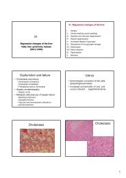
Results: The highest macroscopic lesion was cirrhosis <strong>and</strong> then lesions<br />
due to hydatid cysts <strong>and</strong> Fascioulosis. In microscopic study, the highest<br />
diagnosed lesions was Bacterial hepatitis 20 cases (12.5 %) <strong>and</strong> then<br />
pathological lesions due to Fascioulosis 18 cases (11.35%), hepatic<br />
cirrhosis16 cases(%10), Chronic bill ducts inflammation 16 cases(10%),<br />
Telangiectasia16 cases (10%), Dicroselliousis 14 cases (8.75%), Chronic<br />
hepatitis 14 cases (8.75%), Hydatid cyst 10 cases (6.25%) , Fatty Liver 8<br />
cases (5%),Hemorrhagic Pies hepatitis 8 cases (5%), Blood congestion in<br />
sinusoidal perihepatitis <strong>and</strong> Eosinophilic parasitical hepatitis each of<br />
them 6 cases (3.75%), Cholangitis <strong>and</strong> fibroma 4 cases (2.5%). In this<br />
research, centrilobular Necrosis <strong>and</strong> cholangiocarsinoma which reported<br />
in another spices was none found (0.0%).<br />
Conclusion: The effects of contamination to parasitical agents<br />
identified as the most causes of deletion of sheep liver directly or<br />
indirectly. Even thought the cirrhosis was major factor in macroscopic<br />
judgment for deletion, but the parasitical agents are the essential factors<br />
for deletion of lives.<br />
Key words: sheep health, meat hygiene, pathological lesions of liver,<br />
judgment of carcasses<br />
736 Antibacterial Activity of Aqueous Methanol Extracts of<br />
Zingiber Officianale, Curcuma Longa, Vernonia<br />
Anthelmintica, Acacia Nilotica <strong>and</strong> Melia Azedarach<br />
Z. Abbas 1 , M. Aslam 2 , M. Arshad 2 , Z. Iqba 1 , M. Ashraf 2<br />
1<br />
University of Agriculture, Department of Parasitology, Faisalabad-<br />
38040, Pakistan<br />
2<br />
University of Agriculture, Department of Microbiology, Faisalabad,<br />
Pakistan<br />
Introduction: The development of drug resistance as well as<br />
appearance of undesirable side effects of certain antibiotics has led to<br />
the search of new antibacterial agents in particular from medicinal<br />
plants. Therefore, the present study was conducted to assess the<br />
antibacterial potential of aqueous methanol extract of Zingiber<br />
officianale, Curcuma longa, Vernonia anthelmintica, Acacia nilotica<br />
<strong>and</strong> Melia azedarach against Staphylococcus aureus, Escherichia coli<br />
<strong>and</strong> Bacillus subtilis.<br />
Materials <strong>and</strong> Methods: The antibacterial activity of these extracts<br />
was tested using disc diffusion method. Filter paper discs were soaked<br />
with 15ul of plant extracts stock solution <strong>and</strong> were allowed to dry at<br />
room temperature for 10-15 min. A bacterial culture was uniformly<br />
spread on the surface of Mueller Hinton agar plates using a sterile<br />
swab. Each disc was pressed down uniformly against the surface of<br />
agar <strong>and</strong> then examined for any inhibition zone if present. The test was