Oral and Poster Abstracts
Oral and Poster Abstracts
Oral and Poster Abstracts

Create successful ePaper yourself
Turn your PDF publications into a flip-book with our unique Google optimized e-Paper software.
2<br />
Finnish Food Safety Authority Evira, Veterinary Bacteriology,<br />
Kuopio, Finl<strong>and</strong><br />
3<br />
Finnish Food Safety Authority Evira, Veterinary Virology, Helsinki,<br />
Finl<strong>and</strong><br />
Objectives of the study: Sheep-associated malignant catarrhal fever<br />
(SA-MCF) is a severe <strong>and</strong> mostly fatal disease of cattle <strong>and</strong> other<br />
ruminants. Severe outbreaks have been described in bisons that are the<br />
most susceptible animals. Sheep-associated MCF has been linked to<br />
ovine herpesvirus-2 (OHV-2) infection. We describe here an outbreak<br />
of SA-MCF with high mortality in a Finnish dairy farm.<br />
Materials <strong>and</strong> Methods: The farm. The affected herd consisted of<br />
appr. 40 dairy cows <strong>and</strong> heifers, calves <strong>and</strong> young cattle. All animals<br />
were housed in the same insulated barn with shared air conditioning,<br />
operated with forced exhaust ventilators. Free stall was used for cows,<br />
<strong>and</strong> young cattle were raised in group pens. The farmer brought two<br />
lambs, born in spring 2005, into the dairy barn in November 2005 <strong>and</strong><br />
the lambs stayed in the barn until early April 2006. Examinations:<br />
Pathological examinations were carried out <strong>and</strong> diagnoses were<br />
verified by OHV-2 specific PCR. Serum <strong>and</strong> EDTA blood samples<br />
were collected from all living animals in early April 2006 for analyses<br />
of OHV-2 (PCR) <strong>and</strong> MCF-antibodies (c-ELISA).<br />
Results: In early January 2006 the exhaust ventilators were off for<br />
eight hours due to a power failure. Thus the air quality in the barn was<br />
extremely poor with high humidity <strong>and</strong> severe condensation. First signs<br />
of MCF were seen in February <strong>and</strong> the last case was in November 2006.<br />
The peak of the cases was at spring time. The sings including high<br />
fever, milk drop in cows, hyperaemic oral <strong>and</strong> nasal membranes,<br />
depression, oculo-nasal discharge <strong>and</strong> diarrhoea were seen in a total of<br />
31 animals including cows, calves, heifers <strong>and</strong> young cattle. One cow<br />
with typical clinical signs, high titre of specific antibodies <strong>and</strong> positive<br />
OHV-2 PCR result recovered. The remaining 30 animals with signs of<br />
MCF died or were euthanized. The diagnosis of MCF was based on<br />
gross <strong>and</strong> histopathology <strong>and</strong> presence of both OHV-2 DNA <strong>and</strong><br />
specific antibodies. The animals were also tested for other agents of<br />
upper respiratory tract <strong>and</strong> intestinal infections including respiratory<br />
syncytial virus, infectious bovine rhinotracheitis, bovine coronavirus,<br />
bovine viral diarrhoeal virus, mycoplasmas <strong>and</strong> salmonella, with<br />
negative results.<br />
Conclusions: The adolescent sheep between 6 <strong>and</strong> 9 months of age<br />
have been shown to shed OHV-2 virus more often <strong>and</strong> in greater<br />
amounts than adults. Here keeping of two adolescent sheep in a dairy<br />
barn caused a severe outbreak of MCF in a dairy herd resulting in death<br />
of 30 animals.<br />
662 Evaluation of Nasopharyngeal Swabs in Sampling for<br />
Respiratory Pathogens in Calves<br />
T. Autio 1 , T. Pohjanvirta 2 , A. Huovilainen 3 , L. Sihvonen 3 ,<br />
S. Pelkonen 2<br />
1<br />
Finnish Food Safety Authority Evira, Production Animal Health,<br />
Kuopio, Finl<strong>and</strong><br />
2<br />
Finnish Food Safety Authority Evira, Veterinary Bacteriology,<br />
Kuopio, Finl<strong>and</strong><br />
3<br />
Finnish Food Safety Authority Evira, Veterinary Virology, Helsinki,<br />
Finl<strong>and</strong><br />
Objectives of the study: The aim of this study was to compare<br />
nasopharyngeal swab (NP) <strong>and</strong> bronchoalveolar lavage (BAL) samples<br />
in sampling for respiratory pathogens in calves. The samples were<br />
examined for presence of Pasteurella multocida, bovine coronavirus<br />
(BCV) <strong>and</strong> respiratory syncytial virus (RSV), which are the most<br />
common pathogens associated with calf respiratory disease in Finl<strong>and</strong>.<br />
The similarity of Pasteurella multocida isolates from upper <strong>and</strong> lower<br />
respiratory tract was analysed.<br />
Materials <strong>and</strong> Methods: NP <strong>and</strong> BAL samples were taken in parallel<br />
from 42 calves. NP was taken with a guarded 27-cm swab <strong>and</strong> BAL<br />
with a guarded nasal tube. The samples were examined<br />
bacteriologically by st<strong>and</strong>ard laboratory methods. Pulsed-field gel<br />
electrophoresis (SalI) was utilized for genetic typing of P. multocida<br />
isolates. BCV <strong>and</strong> RSV were detected with RT-PCR.<br />
Results: Five animals were negative for the studied pathogens. Both P.<br />
multocida <strong>and</strong> BCV were detected in 14 animals. A combination of P.<br />
multocida, RSV <strong>and</strong> BCV was found in two animals, <strong>and</strong> three were<br />
infected with both P. multocida <strong>and</strong> RSV. A total of five animals<br />
harboured RSV, of which two in both samples <strong>and</strong> two only in NP.<br />
BCV was detected in nine animals by both sampling techniques <strong>and</strong> in<br />
eight <strong>and</strong> three animals only in NP or BAL, respectively. P. multocida<br />
was detected in 33 animals. Out of these, 23 calves harboured P.<br />
multocida both in NP <strong>and</strong> BAL. All BAL samples positive for P.<br />
multocida were also NP positive, <strong>and</strong> 10 only in NP. Paired P.<br />
multocida isolates obtained from 23 animals with both sampling<br />
methods were characterized by PFGE. Majority of animals (20/23)<br />
harboured isolates showing similar macrorestriction patterns (MRP) or<br />
had minor differences in their MRPs.<br />
Conclusions: Nasopharyngeal swabbing was found to be a suitable<br />
method for sampling for BCV, RSV <strong>and</strong> P. multocida. All pathogens<br />
were recovered more often in nasopharyngeal swab than in BAL<br />
samples. PFGE typing indicated that in most Pasteurella cases the same<br />
strain was isolated by both sampling methods. Thus the P. multocida<br />
culture obtained by nasopharyngeal swabbing represents reasonable<br />
well for the microbes in the lung.<br />
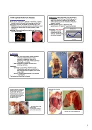
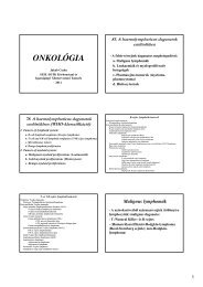
663 First Case of Immunohistochemical Detection of Mycoplasma<br />
bovis in Natural Cases of Calf Pneumonia in Hungary<br />
L. Szeredi 1 , T. Labossa 2 , M. Tenk 3 , V. Pálfi 1 , D. Rigó 1 , T. Molnár 1<br />
1 Central Agricultural Office, Budapest, Hungary<br />
2 Veterinary Practitioner, Lajoskomárom, Hungary<br />
3 CEVA, Budapest, Hungary<br />
Objectives of the study: Bovine respiratory disease complex (BRDC)<br />
is a major cause of morbidity, mortality <strong>and</strong> economic losses in cattle<br />
production systems. The cause of this disease is rather complex<br />
involving both managing factors <strong>and</strong> infectious agents (bacteria <strong>and</strong><br />
viruses). In our study, we demonstrate the primary role of Mycoplasma<br />
(M.) bovis in a severe respiratory disease in feedlot cattle.<br />
Materials <strong>and</strong> methods: Five feedlot cattle died after a few days of<br />
illness without any specific symptom in a herd. Out of these 2 were<br />
treated with antibiotics whereas 3 suddenly died without any treatment.<br />
All the animals were imported from Pol<strong>and</strong>, vaccinated previously<br />
against infectious bovine rhinotracheitis (IBR) <strong>and</strong> bovine respiratory<br />
syntitial virus (BRSV). After gross pathological investigation swabs<br />
were collected for bacterial culture. From the lungs, mycoplasma<br />
culture was also made in 1 case <strong>and</strong> virus isolation (VI) was attempted<br />
in 4 cases. Tissue samples were collected from the lungs <strong>and</strong><br />
occasionally also from the trachea <strong>and</strong> the mediastinal lymph nodes for<br />
histological examination. Immunohistochemical (IHC) method was<br />
used to detect viruses (bovine herpesvirus 1 (BHV-1), BRSV, bovine<br />
respiratory coronavirus, influenza A virus) <strong>and</strong> bacteria (M. bovis <strong>and</strong><br />
Chlamydiaceae) in formalin-fixed paraffin-embedded tissue samples.<br />
Results: The cranioventral part or the whole lungs were affected by<br />
dark discoloration in all cases. Histologically, fibrinopurulent<br />
pleuropneumonia, suppurative bronchiolitis <strong>and</strong> focal to coalescing<br />
coagulative necrosis were observed in the lungs in all cases. M. bovis<br />
was detected in large numbers in the lung tissue samples with IHC,<br />
mostly in the area of bronchiolitis <strong>and</strong> coagulative necrosis, <strong>and</strong> in<br />
smaller number in the area of the fibrinopurulent inflammation. M.<br />
bovis was isolated from one of the cases. Other pathogens were<br />
detected only infrequently: parainfluenza 3 virus (1 case with VI), RSV<br />
(1 case with IHC), Pasteurella multocida (2 cases with culture),<br />
Mannheimia haemolytica (1 case with culture).<br />
Conclusions: M. bovis was the most important pathogenic agent that<br />
induced a severe respiratory disease in the feedlot cattle examined in<br />
this study. Other bacteria <strong>and</strong> viruses were detected only in a few cases.<br />
In spite of the sudden death in 3 cases, the lung lesions were not acute<br />
in any of the cases.<br />
664 Short-duration Antimicrobial Treatment of Bovine<br />
Respiratory Disease with Enrofloxacin, Florfenicol <strong>and</strong><br />
Cefquinome<br />
R. Froyman 1 , C. Boda 2 , C. Rizet 3 , A. Valognes 4 , N. Brunner 1 ,<br />
P. Liege 2 , H. Navetat 3<br />
1 Bayer HealthCare, Animal Health Division, 51368 Leverkusen,<br />
Germany<br />
2 Anisteme Biosciences, Animal Health Division, 34400 Saint-<br />
Christol, France<br />
3 Centre d’Information Veterinaire Clinique, 03130 Le Donjon,<br />
France<br />
4 LDV03, 03017 Moulins, France<br />
Mannheimia haemolytica (Mh) is the most prevalent causative<br />
bacterium in bovine respiratory disease (BRD) but Mycoplasma bovis<br />
Infectious <strong>and</strong> Zoonotic Deseases (Public Health) 91