Oral and Poster Abstracts
Oral and Poster Abstracts
Oral and Poster Abstracts

Create successful ePaper yourself
Turn your PDF publications into a flip-book with our unique Google optimized e-Paper software.
5.00+0.67 <strong>and</strong> 4.94+0.15 mEq/l in the blood plasma of yaks in the<br />
three trimesters of pregnancy, respectively.<br />
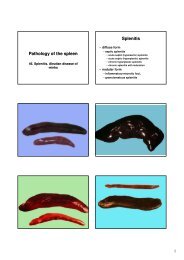
1062 Relationship between Hepatotoxic Compounds <strong>and</strong><br />
Photosensitization in Cattle<br />
J. Seixas 1 , P. Peixoto 2 , C. Pinto 3<br />
1<br />
Universidade Federal Rural do Rio de Janeiro, Curso de Pos<br />
Graduacao em Ciencias Veterinarias, Seropédica, Rio de Janeiro,<br />
Brazil<br />
2<br />
Universidade Federal Rural do Rio de Janeiro, Instituto de<br />
Zootecnia, Seropédica, Rio de Janeiro, Brazil<br />
3<br />
Serviço de Desenvolvimento Agrário de Sao Miguel, Ponta Delgada,<br />
Portugal<br />
Most photosensitization in cattle is caused by phylloerythrin<br />
retention consequential to liver damage caused by hepatotoxic<br />
compounds. Injury to hepatic canalicular membranes <strong>and</strong> subsequent<br />
cholestasis occurs in Lantana spp poisoning, although there is also<br />
significant parenchimal lesions. Primary hepatocellular injury is<br />
caused by the consumption of Myoporum laetum. Photosensitization<br />
caused by sporidesmin from Pithomyces chartarum is characterized<br />
mainly by marked fibrosis biliary obstruction may be caused by the<br />
presence of birefringents crystals. The consumption of plants<br />
containing saponins causes icterus which has, as one of the main<br />
characteristics, crystal-associated cholangiopathy. Poisoning by<br />
saponins is developed after consumption of Tribulus terrestris,<br />
Panicum spp, Narthecium ossifragum <strong>and</strong> Brachiaria spp. This work<br />
describes the main hepatotoxins causing cattle photosensitization<br />
<strong>and</strong> relates them to their pathogenetic effects. The pentacyclic<br />
triterpenoids (present in Lantana spp) cause intrahepatic cholestasis<br />
with the inhibition of bile secretion. The metabolism of essential oils<br />
of furanosesquiterpenes (M. laetum) cause hepatic necrosis. Saponin<br />
poisoning normally occurs with hepatic biliary crystal obstruction.<br />
Injury to the biliary ducts caused by the sporidesmin is characterized<br />
by severe pericholangitis caused by the generation of oxygen free<br />
radicals <strong>and</strong> resulting in fibrosis. There is no scientific consensus<br />
about T. terrestris <strong>and</strong> N. ossifragum poisoning concerning hepatic<br />
target lesions: hepatocellular or biliary system. Brachiaria spp<br />
poisoning should be better included in the group of primary liver<br />
parenchymal damaging plants, because of although it occur, in a<br />
small percentage of cases, deposition of crystals birefringents in the<br />
biliary ducts, those lesions are, in general, enough located. On the<br />
other h<strong>and</strong>, ultramicroscopy reveals marked deposition of crystals<br />
inside hepatocytes, with development of degenerative alterations. It<br />
is also very characteristic the formation of numerous foam cells,<br />
general under form of foci (crystals <strong>and</strong> remainders of necrotic<br />
hepatocytes inside the macrophages) (Lemos et al., 1997; Driemeier<br />
et al., 1998; Gomar et al., 2005; Seixas & Peixoto, 2007). Those<br />
characteristics indicate that the phylloerythrin retention in case of<br />
Brachiaria spp poisoning is doing in the hepatic parenchyma <strong>and</strong> not<br />
in the biliary system.<br />
Key words: photosensitization, hepatotoxic compounds, saponins,<br />
sporidesmin, Brachiaria spp<br />
1063 Urinary Bladder Rupture in Consequence of Urolithiasis in<br />
Nelore Cattle<br />
R. Godoy, L. Gouvea, C. Leite, C. Pereira, FH. Ximenes,<br />
L. Gontijo, R. Ferreira II, JR. Borges<br />
Universidade de Brasília, FAV, Brasília-DF Brazil, Brazil<br />
Objective: The case report is of Nelore cattle, 28 months, male,<br />
presenting obstructive urolithiasis <strong>and</strong> urinary bladder rupture,<br />
examined at the School Hospital of Large Animals at the University of<br />
Brasília.<br />
Material <strong>and</strong> method the animal had anuria, tachycardia,<br />
dehydration, abdominal pain <strong>and</strong> azotemia. It was on exhibition for<br />
24 days, being fed with hay, silage <strong>and</strong> concentrate. Trans-rectal<br />
palpation showed a filled bladder, soon after that the animal expelled<br />
a calculi. The urine had reddish colour, density 1,010, pH 7.0,<br />
protein (++), haemoglobin (++++) <strong>and</strong> macroscopically a lot of<br />
smaller stones could be visualized. The treatment during<br />
hospitalization was of continuous intravascular fluid, vitamin C<br />
(10mg/Kg - TID - 3 days), enrofloxacin (5mg/Kg - SID - 5 days),<br />
flunixim meglumine (1.1 mg / kg - SID - 2 days), oral-ruminal<br />
hydration <strong>and</strong> restricted diet of grass. The improvement was evident<br />
302 XXV. Jubilee World Buiatrics Congress 2008<br />
by the obtention of normal colored urine in the end of the second day<br />
of treatment. The animal was released with guidance of restriction of<br />
continuous concentrate feed as treatment. Forty days after, it<br />
returned to the Veterinary Hospital showing symptoms worsened to<br />
his previous hospitalization, again with a history of beind fed with<br />
concentrate. The animal was apathetic with dirty nostrils, reluctant<br />
to move, tachycardiac, with severe dehydration, tachypnea,<br />
abdominal pain <strong>and</strong> ruminal. Paracentesis revealed large quantities<br />
of fluid with odor resembling urine. The presence of a greater<br />
concentration of creatinine in the peritoneal fluid (42.5 mg / dL)<br />
compared to its serum concentration (10.5 mg / dL) proved the fact<br />
of it being urine. Exploratory laparotomy was performed <strong>and</strong><br />
confirmed the rupture of bladder.<br />
Results: after analysis of laboratory tests, clinical evaluation <strong>and</strong><br />
laparotomy, euthanasia was performed. At necropsy, macroscopically<br />
confirmed the bladder rupture <strong>and</strong> found to be calculi of 2.5 x1, 0cm the<br />
cause of obstruction in urethra near the glans.<br />
Conclusion: It was concluded that treatment done during the first<br />
hospitalization was effective, however, animals with a predisposition<br />
to the formation of calculi that continue to receive food rich in<br />
concentrate <strong>and</strong> are subjected to constant stress, may have recurrent<br />
obstructions. Comparison of the levels of creatinine, in the serum <strong>and</strong><br />
peritoneum, proved to be an important tool in the diagnosis of the<br />
presence of urine in the abdominal cavity.<br />
Key words: calculi, urethra obstruction, creatinine<br />
1064 Carcass pH <strong>and</strong> Temperature of Supply Animals with<br />
Organic Chromium, of Nellore <strong>and</strong> F1 Brangus x Nellore<br />
Cattle on Pasture System<br />
A. Jorge 1 , A. Polizel Neto 1 , P. Moreira 2 , A. Ramos 1 , J. Souza 3 ,<br />
R. Pinheiro 1 , C. Francisco 1<br />
1 UNESP- School of Veterinary Medicine <strong>and</strong> Animal Sciences,<br />
Animal production, Botucatu, Brazil<br />
2 UFMT, Animal Production, Sinop, Brazil<br />
3 UFPR, Animal Science Department, Palotina, Brazil<br />
Chromium works as component integral <strong>and</strong> biologically that<br />
potential the insulin action in the cells, stimulating there of keep the<br />
glycogenic <strong>and</strong> this affecting post mortem pH. The aim of this study<br />
was evaluate the effect of the organic chromium supplementation in<br />
the carcass pH curve <strong>and</strong> freezing, of the two racial groups of steers,<br />
Nellore e F1 Brangus x Nellore, on pasture system. Were used two<br />
groups composed 18 animals each (9 Nellore e 9 F 1 Brangus x<br />
Nellore), submitted to two experimental treatments, with <strong>and</strong><br />
without organic chromium supplementation. The animals were<br />
distributed in the treatments at 210 days until at 600 days of age,<br />
when the animals were slaughed, <strong>and</strong> gauged the pH <strong>and</strong> the<br />
temperature at 2, 10 <strong>and</strong> 24 hours after animals died. Not was<br />
detected racial group, treatment <strong>and</strong> interaction influence to any<br />
characteristics evaluated. The conclusion was that the organic<br />
chromium supplementation to beef cattle, on pasture system, not<br />
influences the carcass pH curve <strong>and</strong> temperature.<br />
Key words: bovine, chromium, meat, minerals, supplementation<br />
1065 Evaluation of Carcass Alteration, by Ultrasound Measures,<br />
of Nellore <strong>and</strong> F1 Brangus x Nellore Steers Supply with<br />
Organic Chromium in Pasture System<br />
A. Jorge 1 , A. Polizel Neto 1 , P. Moreira 2 , A. Ramos 1 , J. Souza 3 ,<br />
R. Pinheiro 1 , C. Francisco 1<br />
1 UNESP School of Veterinary Medicine <strong>and</strong> Animal Sciences, Animal<br />
Science Department, Botucatu, Brazil<br />
2 UFMT, Animal Science Department, Sinop, Brazil<br />
3 UFPR, Animal Science Department, Palotina, Brazil<br />
As chromium is related to insulin, supplementing with bio available<br />
organic molecules containing chromium could be quite important for<br />
improving muscular growth <strong>and</strong> carcass quality. Thus the objective of<br />
this experiment was to evaluate the carcass alterations, by ultrasound<br />
measures of the Nellore <strong>and</strong> F 1 Brangus x Nellore steers supply with<br />
organic chromium, in pasture system. Were used two groups with 18<br />
animals each (9 Nellore <strong>and</strong> 9 F 1 Brangus x Nellore), submitted to two<br />
experimental treatments: with <strong>and</strong> without organic chromium<br />
supplementation. Were measures, in two experimental moments, at 450<br />
(I) <strong>and</strong> 600 (II) days of age, the rib eye area (REA_I <strong>and</strong> REA_II), in<br />
cm 2 ; the sub cutaneus fat thickness at the back <strong>and</strong> at the rump, in mm,