Online proceedings - EDA Publishing Association
Online proceedings - EDA Publishing Association
Online proceedings - EDA Publishing Association

Create successful ePaper yourself
Turn your PDF publications into a flip-book with our unique Google optimized e-Paper software.
dipping method, hydrodynamic transmission of the fluid in a<br />
microenvironment is used to drive the motion of the<br />
biomolecules. Tabeling et al. [10] mentioned that when a fluid<br />
flows through a square-shaped downward concave slot, the<br />
fluid causes a degree of swirling flow in the corner of the slot,<br />
and the side length ratio of the square-shaped slot influences the<br />
flow state of the fluid in the slot. Bruus et al. [11] also observed<br />
that the fluid created a swirling flow in the corner of the<br />
square-shaped slot when the Reynolds number of the<br />
microchannel increased. In this study, we first applied the<br />
special structure present in the microenvironment to cause the<br />
internal fluid to swirl in multiple directions, to increase the<br />
chaos of the fluids in the microenvironment. Next, by the<br />
traction force of the chaotic fluid flow, we drove the motion of<br />
the biomolecules, raised the evenness and the coverage rate of<br />
adhesion of the biomolecules to the sensing field, reducing the<br />
time for adhesion of the biomolecules to the sensing field, and<br />
improving the efficiency and sensibility of the microbial sensor.<br />
Finally, we used the plant virus TYMV to test the effect of the<br />
adhesion of TYMV on the sensing surface of the<br />
microenvironment.<br />
II.<br />
2.1 Sensing Principle<br />
SENSING PRINCIPLE AND SIMULATION<br />
The virus, TYMV, [12] is a tymovirus of the family<br />
Tymoviridae. TYMV are propagated in cabbage leaves and<br />
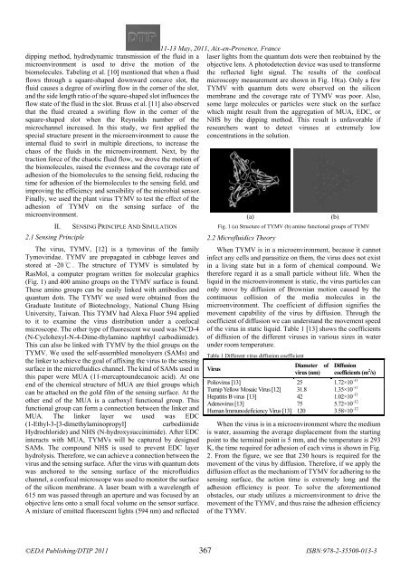
stored at -20 ℃ . The structure of TYMV is simulated by<br />
RasMol, a computer program written for molecular graphics<br />
(Fig. 1) and 400 amino groups on the TYMV surface is found.<br />
These amino groups can be easily linked with antibodies and<br />
quantum dots. The TYMV we used were obtained from the<br />
Graduate Institute of Biotechnology, National Chung Hsing<br />
University, Taiwan. This TYMV had Alexa Fluor 594 applied<br />
to it to examine the virus distribution under a confocal<br />
microscope. The other type of fluorescent we used was NCD-4<br />
(N-Cyclohexyl-N-4-Dime-thylamino naphthyl carbodiimide).<br />
This can also be linked with TYMV by the thiol groups on the<br />
TYMV. We used the self-assembled monolayers (SAMs) and<br />
the linker to achieve the goal of affixing the virus to the sensing<br />
surface in the microfluidics channel. The kind of SAMs used in<br />
this paper were MUA (11-mercaptoundecanoic acid). At one<br />
end of the chemical structure of MUA are thiol groups which<br />
can be attached on the gold film of the sensing surface. At the<br />
other end of the MUA is a carboxyl functional group. This<br />
functional group can form a connection between the linker and<br />
MUA. The linker layer we used was EDC<br />
(1-Ethyl-3-[3-dimethylaminopropyl]<br />
carbodiimide<br />
Hydrochloride) and NHS (N-hydroxysuccinimide). After EDC<br />
interacts with MUA, TYMVs will be captured by designed<br />
SAMs. The compound NHS is used to prevent EDC layer<br />
hydrolysis. Therefore, we can achieve a connection between the<br />
virus and the sensing surface. After the virus with quantum dots<br />
was anchored to the sensing surface of the microfluidics<br />
channel, a confocal microscope was used to monitor the surface<br />
of the silicon membrane. A laser beam with a wavelength of<br />
615 nm was passed through an aperture and was focused by an<br />
objective lens onto a small focal volume on the sensor surface.<br />
A mixture of emitted fluorescent lights (594 nm) and reflected<br />
11-13 <br />
May, 2011, Aix-en-Provence, France<br />
<br />
laser lights from the quantum dots were then reobtained by the<br />
objective lens. A photodetection device was used to transforme<br />
the reflected light signal. The results of the confocal<br />
microscopy measurement are shown in Fig. 10(a). Only a few<br />
TYMV with quantum dots were observed on the silicon<br />
membrane and the coverage rate of TYMV was poor. Also,<br />
some large molecules or particles were stuck on the surface<br />
which might result from the aggregation of MUA, EDC, or<br />
NHS by the dipping method. This result is unfavorable if<br />
researchers want to detect viruses at extremely low<br />
concentrations in the solution.<br />
(a)<br />
(b)<br />
Fig. 1 (a) Structure of TYMV (b) amine functional groups of TYMV<br />
2.2 Microfluidics Theory<br />
When TYMV is in a microenvironment, because it cannot<br />
infect any cells and parasitize on them, the virus does not exist<br />
in a living state but in a form of chemical compound. We<br />
therefore regard it as a small particle without life. When the<br />
liquid in the microenvironment is static, the virus particles can<br />
only move by diffusion of Brownian motion caused by the<br />
continuous collision of the media molecules in the<br />
microenvironment. The coefficient of diffusion signifies the<br />
movement capability of the virus by diffusion. Through the<br />
coefficient of diffusion we can understand the movement speed<br />
of the virus in static liquid. Table 1 [13] shows the coefficients<br />
of diffusion of the different viruses in various sizes in water<br />
under room temperature.<br />
Table 1 Different virus diffusion coefficient<br />
Diameter of<br />
Virus<br />
virus (nm)<br />
Diffusion<br />
coefficients (m 2 /s)<br />
Poliovirus [13] 25 1.72×10 –11<br />
Turnip Yellow Mosaic Virus [12] 31.8 1.35×10 –11<br />
Hepatitis B virus [13] 42 1.02×10 –11<br />
Adenovirus [13] 75 5.72×10 –12<br />
Human Immunodeficiency Virus [13] 120 3.58×10 –12<br />
When the virus is in a microenvironment where the medium<br />
is water, assuming the average displacement from the starting<br />
point to the terminal point is 5 mm, and the temperature is 293<br />
K, the time required for adhesion of each virus is shown in Fig.<br />
2. From the figure, we see that 230 hours is required for the<br />
movement of the virus by diffusion. Therefore, if we apply the<br />
diffusion effect as the mechanism of TYMV for adhering to the<br />
sensing surface, the action time is extremely long and the<br />
adhesion efficiency is poor. To solve the aforementioned<br />
obstacles, our study utilizes a microenvironment to drive the<br />
movement of the TYMV, and thus raise the adhesion efficiency<br />
of the TYMV.<br />
367