Biomechanics and Medicine in Swimming XI
Biomechanics and Medicine in Swimming XI
Biomechanics and Medicine in Swimming XI

You also want an ePaper? Increase the reach of your titles
YUMPU automatically turns print PDFs into web optimized ePapers that Google loves.
Muscle Fatigue <strong>in</strong> Swimm<strong>in</strong>g<br />
rouard, A.h.<br />
Laboratoire de Physiologie de l’Exercice, Université de Savoie, France<br />
Fatigue is a complex process which could be related to different alterations<br />
either of the central nervous system <strong>and</strong>/or of the muscles.<br />
Few studies have focused on the biomechanical evaluation of fatigue<br />
<strong>in</strong> swimm<strong>in</strong>g. EMG results <strong>in</strong>dicated either an <strong>in</strong>crease of <strong>in</strong>tegrated<br />
EMG (IEMG) for muscles with sub-maximal activation or a decrease<br />
of IEMG for muscles strongly <strong>in</strong>volved. Moreover, the frequency contents<br />
shift toward lower frequencies either for Maximal Voluntary Contraction<br />
(MVC) realised before <strong>and</strong> after an exhaustive test or dur<strong>in</strong>g a<br />
maximal swimm<strong>in</strong>g test. Changes <strong>in</strong> muscular activation were associated<br />
with a decrease of force production (dry l<strong>and</strong> strength or tethered or<br />
semi-tethered or swimm<strong>in</strong>g h<strong>and</strong> forces) <strong>and</strong> to changes <strong>in</strong> the path of<br />
the h<strong>and</strong>. Fatigue appears to be related to the task, the subject <strong>and</strong> the<br />
muscle.<br />
Key words: front crawl, fatigue, electromyography, forces, k<strong>in</strong>ematics.<br />
IntroductIon<br />
The swimmers performance is determ<strong>in</strong>ed by the ability to generate<br />
propulsive forces while reduc<strong>in</strong>g the resistance to forward motion. Propulsive<br />
forces are ma<strong>in</strong>ly generated by 3D limb movements <strong>in</strong> response<br />
to unstable loads created by the water. As <strong>in</strong> all human activities, fatigue<br />
could be def<strong>in</strong>ed as an acute impairment of performance. In regard to<br />
the movement generation process, fatigue could be related to central<br />
<strong>and</strong>/or peripheral alterations. The central component of fatigue could be<br />
due to the decrease of the CNS activation (nervous order <strong>and</strong>/ or motor<br />
neuron activation). The peripheral fatigue is related to an alteration<br />
of the neuromuscular junction <strong>and</strong>/or the deficit of substrates, blood<br />
flow, <strong>and</strong>/or dysfunction of the sarcomer. Consequently, fatigue is a very<br />
complex phenomenon, which could be evaluated through different approaches<br />
(physiology, Electromyography (EMG) <strong>and</strong> Mechanics).<br />
Because of the complexity of the movement <strong>in</strong> an aquatic environment,<br />
few biomechanical studies have focused on fatigue <strong>in</strong> swimm<strong>in</strong>g.<br />
EMG has largely been used <strong>in</strong> the evaluation of fatigue dur<strong>in</strong>g susta<strong>in</strong>ed<br />
isometric contractions. Many authors have observed a shift to<br />
lower frequencies of the EMG signal spectrum. In the 90’s, a novel approach<br />
(time-frequency treatment) was proposed for calculat<strong>in</strong>g spectral<br />
parameters from the surface myo-electric signal dur<strong>in</strong>g cyclic dynamic<br />
contractions for which changes <strong>in</strong> muscle length, force <strong>and</strong> electrode<br />
position contributed to the non-stationary status of the signal (Knaflitz<br />
<strong>and</strong> Bonato, 1999). Recently this approach was applied to swimm<strong>in</strong>g<br />
movement (Caty et al, 2007).<br />
The present review concerns the effect of fatigue on muscular activation<br />
<strong>and</strong> the associated changes <strong>in</strong> forces <strong>and</strong> h<strong>and</strong> trajectories<br />
Methods<br />
Most of the studies on fatigue <strong>in</strong> swimm<strong>in</strong>g have been conducted on<br />
male <strong>in</strong>ternational swimmers. Different maximal tests were performed<br />
by the subjects depend<strong>in</strong>g on the authors (i.e. maximal 400m swim <strong>in</strong> a<br />
flume for Monteil et al, 1996, or maximal 4*50m for Caty et al, 2007).<br />
Dur<strong>in</strong>g the test, EMG was synchronised with video acquisition. For<br />
EMG, the authors used either surface electrodes (Wakayoshi et al, 1994,<br />
Rouard et al, 1997, Caty et al, 2007) or f<strong>in</strong>e wire electrodes (Monteil et al,<br />
1996). Shoulder <strong>and</strong> upper limb muscles were those most <strong>in</strong>vestigated.<br />
All the authors applied the same guidel<strong>in</strong>es for the location of the electrodes<br />
(belly of the muscle) <strong>and</strong> the sk<strong>in</strong> preparation (Clarys <strong>and</strong> Cabri,<br />
1993). Signals were stored on a memory card or on the soundtrack of the<br />
video camera. For the amplitude treatment, the raw EMG signals were<br />
rectified, smoothed <strong>and</strong> then <strong>in</strong>tegrated (IEMG). For each subject, <strong>and</strong><br />
each muscle, the IEMG were normalised accord<strong>in</strong>g to the maximal dy-<br />
chaPter1.<strong>in</strong>vitedLectures<br />
namic value observed dur<strong>in</strong>g the test<strong>in</strong>g procedures. For the frequency<br />
treatment, Aujouannet et al (2006) applied a Fourier transformation to<br />
evaluate the frequency contents of static Maximal Voluntary Contractions<br />
(MVC) performed just before <strong>and</strong> after the exhaustive swimm<strong>in</strong>g<br />
test (4*50m). For the swimm<strong>in</strong>g signals, Caty et al (2007) extracted, with<br />
a statistical detector, the activation <strong>in</strong>terval correspond<strong>in</strong>g to each stroke<br />
<strong>and</strong> each muscle. For each detected muscle burst, the Choi-Williams<br />
transformation was then computed. These particular transformations<br />
had already proven effective <strong>in</strong> the analysis of strongly non-stationary<br />
EMG signals recorded dur<strong>in</strong>g dynamic exercise (Knaflitz <strong>and</strong> Bonato,<br />
1999). The <strong>in</strong>stantaneous mean frequency of the signal burst (MNF, Hz)<br />
was calculated for each stroke cycle.<br />
For the k<strong>in</strong>ematic evaluation of fatigue, at least 2 underwater cameras<br />
were used to analyse the 3D movements of the upper limbs. The<br />
video was digitised frame by frame to get the h<strong>and</strong> trajectories <strong>and</strong>/or<br />
the h<strong>and</strong> velocity <strong>and</strong>/or different angles (sweep back <strong>and</strong> attack h<strong>and</strong><br />
angles, limbs angles).<br />
Dur<strong>in</strong>g the MVC, the forces were recorded us<strong>in</strong>g a stra<strong>in</strong> gauge, while<br />
dur<strong>in</strong>g swimm<strong>in</strong>g the authors used either direct force measurements<br />
such as a tethered or semi- tethered apparatus (Aujouannet et al, 2006,<br />
Rouard et al, 2006) or an <strong>in</strong>direct approach calculat<strong>in</strong>g the lift, drag<br />
<strong>and</strong> resultant h<strong>and</strong> forces accord<strong>in</strong>g to Schleihauf ’s method (Monteil<br />
et al, 1996)<br />
results<br />
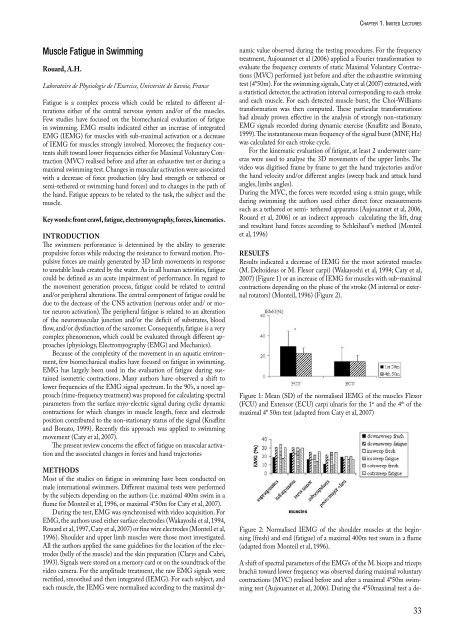
Results <strong>in</strong>dicated a decrease of IEMG for the most activated muscles<br />
(M. Deltoideus or M. Flexor carpi) (Wakayoshi et al, 1994; Caty et al,<br />
2007) (Figure 1) or an <strong>in</strong>crease of IEMG for muscles with sub-maximal<br />
contractions depend<strong>in</strong>g on the phase of the stroke (M <strong>in</strong>ternal or external<br />
rotators) (Monteil, 1996) (Figure 2).<br />
Figure 1: Mean (SD) of the normalised IEMG of the muscles Flexor<br />
(FCU) <strong>and</strong> Extensor (ECU) carpi ulnaris for the 1 st <strong>and</strong> the 4 th of the<br />
maximal 4* 50m test (adapted from Caty et al, 2007)<br />
Figure 2: Normalised IEMG of the shoulder muscles at the beg<strong>in</strong>n<strong>in</strong>g<br />
(fresh) <strong>and</strong> end (fatigue) of a maximal 400m test swam <strong>in</strong> a flume<br />
(adapted from Monteil et al, 1996).<br />
A shift of spectral parameters of the EMG’s of the M. biceps <strong>and</strong> triceps<br />
brachii toward lower frequency was observed dur<strong>in</strong>g maximal voluntary<br />
contractions (MVC) realised before <strong>and</strong> after a maximal 4*50m swimm<strong>in</strong>g<br />
test (Aujouannet et al, 2006). Dur<strong>in</strong>g the 4*50maximal test a de-<br />
33