

Pathologica 4-07.pdf - Pacini Editore
Pathologica 4-07.pdf - Pacini Editore
Pathologica 4-07.pdf - Pacini Editore

You also want an ePaper? Increase the reach of your titles
YUMPU automatically turns print PDFs into web optimized ePapers that Google loves.
268<br />
GCET1 expression in endemic Burkitt<br />
lymphoma. Correlation with EBV status and<br />
IGH mutation pattern<br />
C. Bellan, S. Lazzi, M. Cocco, T. Amato, N. Palummo, G.<br />
De Falco, E. Leucci, S. Mannucci, P. Tosi, M. Piris * , L.<br />
Leoncini<br />
Dipartimento di Patologia Umana ed Oncologia, Università<br />
di Siena, Italy; * Centro Nacional de Investigaciones Oncologicas<br />
(CNIO), Madrid, Spain<br />
Introduction. GCET1 (centerin, serpin A9) is a gene induced<br />
in B cells by CD40-CD40L interaction and suspected<br />
to play an important role in GC-B cell development 1 . Previous<br />
studies have shown that expression of GCET1 is primarily<br />
restricted to GC B-cells (centroblasts and large centrocytes<br />
but not small centrocytes). The normal counterpart of<br />
the neoplastic B cells in Burkitt lymphoma (BL) is still unclear.<br />
Basing on immunoglobulin gene rearrangement studies,<br />
some authors suggest an origin from germinal center B<br />
cells and others from memory B cells 2 . To better clarify the<br />
cells of origin in BL we analysed GCET1 expression on 38<br />
endemic BL, and we correlated its expression with EBV status<br />
and immunoglobulin gene mutation pattern of these cases.<br />
Materials and methods. All cases were classified as eBLs according<br />
to WHO criterias. IHC for GCET1 expression and ISH<br />
for EBERs were performed on consecutive sections according<br />
to the manufacturers. Groups of 5-7 GCET1 positive cells were<br />
taken through LCM. Following overnight digestion, the lysate<br />
was directly used as a template in a semi-nested PCR amplification<br />
for VH gene rearrangements analysis. The PCR products<br />
were subsequently cloned and sequenced in both directions.<br />
Only sequences showing a perfect homology were chosen for<br />
comparison with germline sequences from ImmunoGeneTiCs<br />
database. V H genes were considered mutated if they differed<br />
2% or more from the corresponding germ line sequence.<br />
Results. We found that GCET1 expression was significantly<br />
correlated with EBV status. In fact, of 31 EBV positive eBLs,<br />
29 did not shown evidence of GCET1 expression, while 5 out<br />
of seven EBV negative eBLs actively expressed the gene.<br />
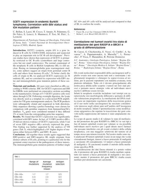
This finding was further supported by the analisys of VH<br />
genes (Tab. I), which highlighted a A9- higher degree of mutation<br />
either between EBV+ and EBV- B cases.<br />
Conclusions. All together these results again suggested that<br />
EBV-positive and EBV-negative BL might originate from<br />
distinct subsets of B cells, pointing to a particular role for the<br />
germinal center reaction in the pathogenesis of these tumors.<br />
The immunoglobulin gene mutation patterns of further single<br />
Tab. I.<br />
Range of Average Antigen<br />
mutations mutation selection<br />
frequency<br />
(mean)<br />
EBV-/A9+ 1-7 1.7 0/5<br />
EBV-/A9- 6-9 2.9 0/2<br />
EBV+/A9+ 7-8 3.1 0/2<br />
EBV+/A9- 5-25 5.1 9/29<br />
GC A9+ and A9- cells will be analysed and compared to that<br />
of BL to confirm the results.<br />
References<br />
1 Frazer JK, et al. Eur J Immunol 2000;30:3039-48.<br />
2 Bellan C, et al. Blood 2005;106:1031-6.<br />
POSTERS<br />
Correlazione nei tumori ovarici tra stato di<br />
metilazione dei geni RASSF1A e BRCA1 e<br />
grado di differenziazione<br />
M. Carosi, G. Chichierchia, E. Vizza * , G. Cutillo * , A. Savarese<br />
** , A. Papatantonakis, A. Marsella *** , L. Perracchio<br />
* , P. Visca, F. Marandino, R. Perrone Donnorso<br />
S.C. Anatomia e Istologia Patologica, Istituto “Regina Elena”,<br />
Roma; * Ginecologia Oncologica, Istituto “Regina Elena”,<br />
Roma; ** Oncologia Medica A, Istituto “Regina Elena”,<br />
Roma; *** Radiologia Istituto “Regina Elena”, Roma<br />
Gli eventi molecolari responsabili della carcinogenesi nell’epitelio<br />
ovario non sono ancora tutti noti e nonostante i miglioramenti<br />
terapeutici e chirurgici la sopravvivenza al termine,<br />
per le pazienti soprattutto con malattia avanzata, resta<br />
piuttosto deludente. Tutto ciò è dovuto principalmente alla<br />
nostra poca abilità nello scoprire i tumori nei loro stati precoci<br />
e pertanto nuove strategie volte ad individuare nuovi<br />
markers debbono essere trovate.<br />
Infatti le neoplasie ovariche includono vari istotipi con caratteristiche<br />
isto-morfologiche differenti e pertanto di difficile<br />
valutazione prognostica. La metilazione del DNA risulta<br />
essere un importante regolatore della trascrizione genica<br />
ed il suo ruolo nella carcinogenesi ha suscitato considerevole<br />
interesse negli ultimi anni nell’ambito dell’insorgenza<br />
di molte neoplasie. Infatti l’ipermetilazione come modificazione<br />
epigenetica che reprime la trascrizione dei geni coinvolti<br />
nella soppressione del tumore è stato ampiamente studiato.<br />
Lo scopo di questo studio è di valutare lo stato di metilazione<br />
del promotore dei geni RASSF1A e BRCA1 in vari tipi e<br />
fasi di tumori epiteliali ovarici per valutare se sia possibile<br />
individuare nuovi markers per i quali ideare nuovi farmaci<br />
che possano interferire con gli eventi evolutivi della cellula<br />
neoplastica, con una maggiore selettività del tumore ed al<br />
tempo stesso una minore tossicità. Nel nostro studio sono stati<br />
analizzati 40 tumori epiteliali ovarici in un range che va dai<br />
cistoadenomi benigni, tumori a basso potenziale di malignità<br />
e carcinomi (10 cistoadenomi; 12 tumori a basso potenziale<br />
di malignità e 18 carcinomi) usando il metodo della metilazione-specifica<br />
PCR.<br />
In considerazione dei dati finora ottenuti si e potuto vedere<br />
che il promotore del gene RASSF1A risulta metilato soprattutto<br />
nei tumori a basso potenziale di malignità e nei carcinomi;<br />
mentre il promotore del gene BRCA1 risulta essere<br />
metilato prevalentemente nei carcinomi.


