

Labelling Review row-Online
Labelling Review row-Online
Labelling Review row-Online

You also want an ePaper? Increase the reach of your titles
YUMPU automatically turns print PDFs into web optimized ePapers that Google loves.
Deleted in Colorectal Cancer Protein<br />
Clone DM51<br />
1 mL lyophilized NCL-DCC P (HIER)<br />
The deleted in colorectal cancer (DCC) gene located on chromosome 18 is a<br />
tumor suppressor gene that encodes a transmembrane protein structurally<br />
similar to NCAM. The highest reported expression of this protein can be<br />
found in axons of the central and peripheral nervous systems where it<br />
functions as a netrin receptor required for the guidance of the developing<br />
axons. The DCC gene is reported to be expressed in most epithelial tissues<br />
where the protein may participate in the regulation of cell to cell or cell to<br />
substratum interaction. In normal colon, DCC expression is restricted to the<br />
mucosa with intense granular cytoplasmic staining in the crypts, particularly<br />
in the goblet cells. Altered DCC expression may be the result of allelic loss<br />
which is reported to occur in more than 70 percent of colorectal<br />
carcinomas, localized mutations, aberrant splicing of transcripts or allelespecific<br />
loss of transcripts. The DCC gene has also been reported to be<br />
inactivated in pancreatic, gastric, breast, prostatic and brain cancers and<br />
also in some leukemias. The expression of DCC protein is reduced in these<br />
cancers by 36 to 50 percent. In astrocytic tumors and colorectal carcinomas<br />
reduced expression of DCC protein is reported.<br />
Human colonic adenocarcinoma: immunohistochemical staining for deleted in colorectal<br />
cancer protein using NCL-DCC. Note granular cytoplasmic staining of malignant epithelial<br />
cells. Paraffin section.<br />
Deleted in Pancreatic Cancer Locus 4<br />
Protein<br />
Clone JM56<br />
1 mL lyophilized NCL-DPC4 P (HIER)<br />
Deleted in pancreatic cancer locus 4 (DPC4) is a tumor suppressor gene<br />
reported to be frequently mutated or deleted in pancreatic and metastatic<br />
colon cancers. DPC4, also known as Smad4, acts as a cofactor that binds<br />
transforming g<strong>row</strong>th factor-beta receptor-activated Smad2 and Smad3<br />
generating transcriptional complexes which translocate to the nucleus to<br />
participate in sequence-specific DNA-binding and transcriptional activation.<br />
Mutation or deletion of the DPC4 gene is reported in 50 percent of<br />
pancreatic ductal adenocarcinomas and a subset of acute myelogenous<br />
leukemias, biliary tract carcinomas, ovarian, colon and breast cancers. The<br />
expression of DPC4 protein has been reported to be a sensitive and specific<br />
marker for DPC4 gene alterations in pancreatic carcinomas. Loss of DPC4<br />
expression occurs late in the neoplastic progression which leads to the<br />
development of infiltrating pancreatic cancer when it is histologically<br />
recognizable as a carcinoma. The continued expression of DPC4 protein is<br />
reported in pancreatic intraductal papillary mucinous neoplasms (IPMN)<br />
and suggests genetic differences in tumorigenesis from ductal carcinomas.<br />
Desmin<br />
Clone DE-R-11<br />
1 mL, 0.1 mL lyophilized NCL-DES-DERII F P (Enzyme) W<br />
1 mL liquid NCL-L-DES-DERII F P (Enzyme) W<br />
7 mL ready-to-use RTU-DES-DERII F P (Enzyme)<br />
7 mL Bond ready-to-use PA0032 P (HIER)<br />
Product Specific Information<br />
NCL-DES-DERII reacts with an 18 kD rod piece of the intermediate filament<br />
protein desmin (53 kD) in muscle cells. The antibody does not appear to<br />
recognize other intermediate filament proteins. In normal tissues, Clone DE-<br />
RII reacts with both striated (skeletal and cardiac) and smooth muscle cells.<br />
The labeling is confined to the Z bands in skeletal and cardiac muscle giving<br />
a characteristic striated appearance.<br />
Refer to page 27 for the Bond ready-to-use format.<br />
Normal human small intestine: immunohistochemical staining for desmin using<br />
NCL-DES-DERII. Note cytoplasmic staining of muscle cells in the muscularis externa.<br />
Paraffin section.<br />
DOG-1<br />
Clone K9<br />
1 mL, 0.1 mL liquid NCL-L-DOG-1 P (HIER)<br />
7 mL Bond ready-to-use PA0219 P (HIER)<br />
Reference Range<br />
DOG-1, a 986 amino acid protein of unknown function, is expressed<br />
predominantly on the plasma membrane of gastrointestinal stromal tumors<br />
(GISTs) and is rarely expressed in other soft tissue tumors, which, due to<br />
appearance, can be confused with GISTs. Immunoreactivity for DOG-1 has<br />
been reported to be found in 97.8 percent of scorable GISTs, including all KIT<br />
negative GISTs. Reactivity for DOG-1 has been suggested to aid in the<br />
identification of GISTs, including Platelet-Derived G<strong>row</strong>th Factor Receptor<br />
Alpha mutants that fail to express KIT antigen.<br />
Product Specific Information<br />
The use of PBS-based diluents may result in increased background staining.<br />
Refer to page 27 for the Bond ready-to-use format.<br />
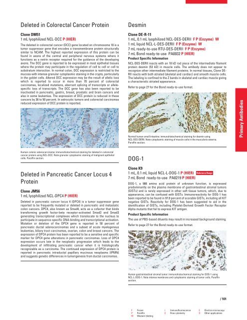
Human gastrointestinal stromal tumor: immunohistochemical staining for DOG-1 using<br />
NCL-L-DOG-1. Note intense membrane and cytoplasmic staining of tumor cells. Paraffin<br />
section.<br />
F Frozen I Immunofluorescence E Electron microscopy<br />
P Paraffin C Flow cytometry O Other applications<br />
W Western blotting<br />
/ 101<br />
Primary Antibodies


