

Labelling Review row-Online
Labelling Review row-Online
Labelling Review row-Online

Create successful ePaper yourself
Turn your PDF publications into a flip-book with our unique Google optimized e-Paper software.
Primary Antibodies<br />
Immunoglobulin D<br />
Clone DRN1C<br />
1 mL, 0.1 mL liquid NCL-L-IgD P (HIER) Reference Range<br />
Clone DRN1C was developed to produce reduced background staining that is<br />
associated with polyclonal antibodies on paraffin sections.<br />
IgD, together with IgM, are the major immunoglobulins expressed on the<br />
surface of B cells where it seems they may operate as mutually interacting<br />
antigen receptors for the control of lymphocyte activation and suppression.<br />
The greater susceptibility of IgD to proteolysis in combination with antigen<br />
could well be implicated in such a function.<br />
Product Specific Information<br />
The use of PBS-based diluents may result in increased background staining.<br />
Clone DRN1C was developed to produce reduced background staining that is<br />
associated with polyclonal antibodies on paraffin sections.<br />
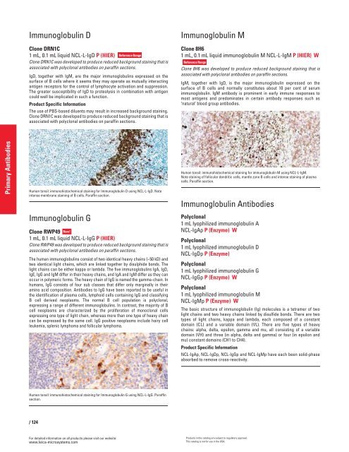
Human tonsil: immunohistochemical staining for Immunoglobulin D using NCL-L-IgD. Note<br />
intense membrane staining of B cells. Paraffin section.<br />
Immunoglobulin G<br />
Clone RWP49 New!<br />
1 mL, 0.1 mL liquid NCL-L-IgG P (HIER)<br />
Clone RWP49 was developed to produce reduced background staining that is<br />
associated with polyclonal antibodies on paraffin sections.<br />
The human immunoglobulins consist of two identical heavy chains (~50 kD) and<br />
two identical light chains, which are linked together by disulphide bonds. The<br />
light chains can be either kappa or lambda. The five immunoglobulins IgA, IgD,<br />
IgE, IgG and IgM differ in their heavy chains, and IgA and IgM differ as they can<br />
occur in polymeric forms. The heavy chain of IgG is named the gamma-chain. In<br />
humans, IgG consists of four sub classes that differ only marginally in their<br />
amino acid composition. Antibodies to IgG have been reported to be useful in<br />
the identification of plasma cells, lymphoid cells containing IgG and classifying<br />
B cell derived neoplasms. The normal B cell population is polyclonal,<br />
expressing a range of different immunoglobulins. In contrast, the majority of B<br />
cell neoplasms are characterized by the proliferation of monoclonal cells<br />
expressing one type of light chain, whereas more than one type of heavy chain<br />
can be expressed by the same cell. IgG positive neoplasms include hairy cell<br />
leukemia, splenic lymphoma and follicular lymphoma.<br />
Human tonsil: immunohistochemical staining for Immunoglobulin G using NCL-L-IgG. Paraffin<br />
section.<br />
/ 124<br />
For detailed information on all products please visit our website:<br />
www.leica-microsystems.com<br />
Immunoglobulin M<br />
Clone 8H6<br />
1 mL, 0.1 mL liquid immunoglobulin M NCL-L-IgM P (HIER) W<br />
Reference Range<br />
Clone 8H6 was developed to produce reduced background staining that is<br />
associated with polyclonal antibodies on paraffin sections.<br />
IgM, together with IgD, is the major immunoglobulin expressed on the<br />
surface of B cells and normally constitutes about 10 per cent of serum<br />
immunoglobulin. IgM antibody is prominent in early immune responses to<br />
most antigens and predominates in certain antibody responses such as<br />
‘natural’ blood group antibodies.<br />
Human tonsil: immunohistochemical staining for immunoglobulin M using NCL-L-IgM.<br />
Note staining of follicular dendritic cells, mantle zone B cells and intense staining of plasma<br />
cells. Paraffin section.<br />
Immunoglobulin Antibodies<br />
Polyclonal<br />
1 mL lyophilized immunoglobulin A<br />
NCL-IgAp P (Enzyme) W<br />
Polyclonal<br />
1 mL lyophilized immunoglobulin D<br />
NCL-IgDp P (Enzyme)<br />
Polyclonal<br />
1 mL lyophilized immunoglobulin G<br />
NCL-IgGp P (Enzyme) W<br />
Polyclonal<br />
1 mL lyophilized immunoglobulin M<br />
NCL-IgMp P (Enzyme) W<br />
The basic structure of immunoglobulin (Ig) molecules is a tetramer of two<br />
light chains and two heavy chains linked by disulfide bonds. There are two<br />
types of light chains, kappa and lambda, each composed of a constant<br />
domain (CL) and a variable domain (VL). There are five types of heavy<br />
chains: alpha, delta, epsilon, gamma and mu, all consisting of a variable<br />
domain (VH) and three (in alpha, delta and gamma) or four (in epsilon and<br />
mu) constant domains (CH1 to CH4).<br />
Product Specific Information<br />
NCL-IgAp, NCL-IgDp, NCL-IgGp and NCL-IgMp have each been solid-phase<br />
absorbed to remove cross-reactivity.<br />
Products in this catalog are subject to regulatory approval.<br />
This catalog is not for use in the USA.


