Labelling Review row-Online
Labelling Review row-Online
Labelling Review row-Online
Create successful ePaper yourself
Turn your PDF publications into a flip-book with our unique Google optimized e-Paper software.
Primary Antibodies<br />
MDM2 Protein<br />
Clone 1B10<br />
1 mL, 0.1 mL lyophilized NCL-MDM2 F P (HIER)<br />
The human phosphoprotein homolog of the murine double minute 2 (MDM2)<br />
gene, with a molecular weight of 90 kD (p90), forms a complex with both<br />
mutant and wild type p53 protein. The MDM2 gene product interacts with<br />
p53 protein inhibiting p53-mediated transactivation. Overexpression of<br />
MDM2 overcomes wild type p53 mediated suppression of transformed cell<br />
g<strong>row</strong>th. MDM2 amplification is reported to be observed in some soft tissue<br />
sarcomas, osteosarcomas and high grade malignant gliomas.<br />
Product Specific Information<br />
NCL-MDM2 reacts with the human homolog of MDM2.<br />
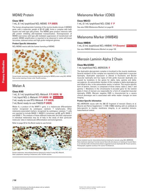
Human breast carcinoma: immunohistochemical staining for MDM2 protein using NCL-MDM2.<br />
Note nuclear staining of tumor cells. Paraffin sectiion.<br />
Melan A<br />
Clone A103<br />
1 mL, 0.1 mL lyophilized NCL-MelanA F P (HIER) W<br />
1 mL liquid NCL-L-MelanA F P (HIER) W<br />
7 mL ready-to-use RTU-MelanA F P (HIER)<br />
7 mL Bond ready-to-use PA0233 P (HIER)<br />
Melan A, a product of the MART-1 gene, is a melanocyte differentiation<br />
marker recognized by autologous cytotoxic T lymphocytes. Other<br />
melanoma-associated markers recognized by autologous cytotoxic T cells<br />
are reported to include MAGE-1, MAGE-3, tyrosinase, gp100, gp75, BAGE-1<br />
and GAGE-1. The analysis of these different molecules and their expression<br />
in individual melanomas may be of help in the study of their particular<br />
molecular roles in melanocyte differentiation and tumorigenesis.<br />
Refer to page 33 for the Bond ready-to-use format.<br />
Human melanoma: immunohistochemical staining for melan A using NCL-MelanA.<br />
Note cytoplasmic staining of melanoma cells. Paraffin section.<br />
/ 132<br />
For detailed information on all products please visit our website:<br />
www.leica-microsystems.com<br />
Reference Range<br />
Melanoma Marker (CD63)<br />
Clone NKI/C3<br />
1 mL, 0.1 mL lyophilized NCL-CD63 FP<br />
See also CD63 (Melanoma Marker) on page 82.<br />
Melanoma Marker (HMB45)<br />
Clone HMB45<br />
1 mL, 0.1mL lyophilized NCL-HMB45 F P (Enzyme)<br />
See also HMB45 (Melanoma Marker) on page 120.<br />
Merosin Laminin Alpha 2 Chain<br />
Clone Mer3/22B2<br />
1 mL lyophilized NCL-MEROSIN F<br />
The dystrophin-glycoprotein complex is localized to the muscle membrane.<br />
Several members of this complex are reported to be implicated in muscular<br />
dystrophy. Dystrophin expression is altered in Duchenne and Becker<br />
muscular dystrophy and four types of limb girdle muscular dystrophy are<br />
caused by mutations in the genes for alpha, beta, gamma and deltasarcoglycan.<br />
An extracellular member of this complex is alpha-dystroglycan<br />
and linked to this, in the extracellular matrix, is laminin. The muscle specific<br />
form of laminin, merosin, is composed of three chains: alpha 2, beta 1 and<br />
gamma 1. Mutations in the chromosome 6 encoded gene for the laminin<br />
alpha 2 chain of merosin are responsible for a form of congenital muscular<br />
dystrophy (CMD). Merosin negative CMD is characterized by a severe<br />
clinical phenotype and is associated with white matter changes on brain<br />
imaging.<br />
Product Specific Information<br />
NCL-MEROSIN reacts with the 300 kD fragment of merosin (Sewry et al.<br />
Muscle and Nerve Supplement. 7, S109: (1998)) labeling with an antibody to<br />
beta-spectrin to monitor membrane integrity, is an essential immunohistochemical<br />
control.<br />
A B<br />
Human skeletal muscle: immunohistochemical staining for merosin using NCL-MEROSIN.<br />
Note membrane staining of normal muscle fibers (A) and absence of staining of muscle fibers<br />
in an individual with chromosome 6-linked congenital muscular dystrophy (B). Frozen sections.<br />
Photographs supplied courtesy of Dr Louise V B Anderson.<br />
Products in this catalog are subject to regulatory approval.<br />
This catalog is not for use in the USA.<br />
Reference Range


