

Labelling Review row-Online
Labelling Review row-Online
Labelling Review row-Online

You also want an ePaper? Increase the reach of your titles
YUMPU automatically turns print PDFs into web optimized ePapers that Google loves.
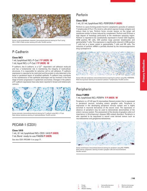
Human acute lymphoblastic leukemia: immunohistochemical staining for Pax-5 using<br />
NCL-L-PAX-5. Note nuclear staining of B cells. Paraffin section.<br />
P-Cadherin<br />
Clone 56C1<br />
1 mL lyophilized NCL-P-Cad F P (HIER) W<br />
1 mL liquid NCL-L-P-Cad F P (HIER) W<br />
P-cadherin, like E-cadherin, is a Ca 2+ -dependent cell adhesion molecule<br />
and has a fundamental role in maintaining the integrity of multicellular<br />
structures. It is responsible for selective cell to cell adhesion. P-cadherin<br />
expression is reported to be restricted and the protein is only detected in the<br />
basal or parabasal layers of stratified epithelia. P-cadherin may contribute<br />
to the maintenance of the epithelial phenotype and be involved in the final<br />
stage of tumor progression in epidermal carcinomas. Changes in the pattern<br />
of P-cadherin expression have also been reported in breast and melanocytic<br />
cancers.<br />
Human placenta: immunohistochemical staining for P-cadherin using NCL-L-P-Cad.<br />
Note intense membrane staining of cytotrophoblasts. Paraffin section.<br />
PECAM-1 (CD31)<br />
Clone 1A10<br />
1 mL, 0.1 mL lyophilized NCL-CD31-1A10 P (HIER)<br />
7 mL Bond ready-to-use PA0250 P (HIER)<br />
See also CD31 (PECAM-1) on page 77.<br />
Perforin<br />
Clone 5B10<br />
1 mL, 0.1 mL lyophilized NCL-PERFORIN P (HIER)<br />
Perforin is a pore-forming protein found in cytoplasmic granules of cytotoxic<br />
T-lymphocytes (CTLs). CTLs bind to cells which express foreign antigens and<br />
induce them to lyse. Perforin forms circular lesions on the target cell<br />
membrane similar to those induced by complement. Perforin and C9 share a<br />
high degree of homology particularly at the membrane spanning region.<br />
Perforin is reported to be constitutively expressed in human CD3 negative,<br />
CD56 positive NK cells, CD3 positive large granular lymphocytes and<br />
gamma/delta T cells. This expression is significantly induced in CD8 positive<br />
T cells but to a lesser extent in gamma/delta T cells and NK cells. The<br />
induction of perforin mRNA is partially blocked by the immunosuppressive<br />
drug cyclosporin A.<br />
Human follicular lymphoma: immunohistochemical staining for perforin using NCL-PERFORIN.<br />
Note focal granular staining of occasional cytotoxic T lymphocytes. Paraffin section.<br />
Peripherin<br />
Clone PJM50<br />
1 mL lyophilized NCL-PERIPH F P (HIER) W<br />
Peripherin is a 57 kD type III intermediate filament protein that is expressed<br />
in peripheral neurons, including enteric ganglion cells. Peripherin is<br />
expressed in the developing peripheral nervous system and is highly<br />
enriched in neuronal derivatives of the neural crest. The expression or<br />
absence of peripherin may be used to demonstrate abnormalities of the<br />
enteric nervous system. The assessment of the density of ganglion cells is of<br />
importance in Hirschsprung's disease (HD)-related disorders. Peripherin is<br />
also reported to be expressed in neural crest derived tumors such as<br />
neuroblastomas and ganglioneuroblastomas.<br />
Human small bowel: immunohistochemical staining for peripherin using NCL-PERIPH.<br />
Note intense cytoplasmic staining of enteric ganglion cells and neural elements.<br />
Paraffin section.<br />
F Frozen I Immunofluorescence E Electron microscopy<br />
P Paraffin C Flow cytometry O Other applications<br />
W Western blotting<br />
/ 149<br />
Primary Antibodies


