

Labelling Review row-Online
Labelling Review row-Online
Labelling Review row-Online

Create successful ePaper yourself
Turn your PDF publications into a flip-book with our unique Google optimized e-Paper software.
Primary Antibodies<br />
CD35<br />
Clone RLB25<br />
1 mL, 0.1 mL lyophilized NCL-CD35 F P (Enzyme)<br />
The CD35 antigen, also known as CR1 or C3b/C4b R, is a transmembrane<br />
protein of 160 to 250 kD which binds complement components C3b and C4b.<br />
It mediates phagocytosis by neutrophils and monocytes of particles coated<br />
with C3b or C4b. CD35 antigen has an inhibitory effect on complement<br />
activation by both the classical and alternative pathways. CD35 antigen is<br />
reported to be found on erythrocytes, B cells, a subset of T cells, monocytes,<br />
macrophages cultured in vitro, neutrophils, eosinophils, glomerular<br />
podocytes and follicular dendritic cells. Decreased levels of CD35 antigen<br />
has been reported on B cells in patients with HIV infection.<br />
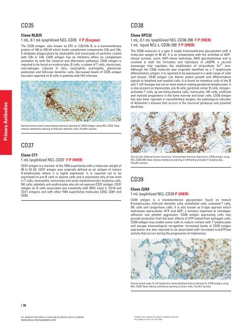
Normal human tonsil: immunohistochemical staining for CD35 antigen using NCL-CD35. Note<br />
intense membrane staining of follicular dendritic cells. Paraffin section.<br />
CD37<br />
Clone CT1<br />
1 mL lyophilized NCL-CD37 F P (HIER)<br />
CD37 antigen is a member of the TM4 superfamily with a molecular weight of<br />
40 to 52 kD. CD37 antigen was originally defined as an antigen of mature<br />
B lymphocytes where it is highly expressed. It is reported not to be<br />
expressed on pre-B cells or plasma cells and is expressed only at low level<br />
in T cells, neutrophils, monocytes and some myelomonocytic leukemia cells.<br />
NK cells, platelets and erythrocytes also do not express CD37 antigen. CD37<br />
antigen on B cells associates non-covalently with MHC class II, CD19 and<br />
CD21 antigens and with other TM4 superfamily molecules CD53, CD81 and<br />
CD82.<br />
/78<br />
For detailed information on all products please visit our website:<br />
www.leica-microsystems.com<br />
CD38<br />
Clone SPC32<br />
1 mL, 0.1 mL lyophilized NCL-CD38-290 F P (HIER)<br />
1 mL liquid NCL-L-CD38-290 F P (HIER)<br />
The CD38 molecule is a type II single transmembrane glycoprotein with a<br />
molecular weight of 46 kD. It is an ectoenzyme with the activities of ADPribosyl<br />
cyclase, cyclic ADP-ribose hydrolase, NAD glycohydrolase and is<br />
involved in both the formation and hydrolysis of cADPR, a second<br />
messenger that regulates the mobilization of intracellular Ca 2+ ions.<br />
Although the CD38 molecule was originally identified as a T lymphocyte<br />
differentiation antigen, it is reported to be expressed in a wide range of cells<br />
and tissues. CD38 antigen can deliver potent g<strong>row</strong>th and differentiation<br />
signals to lymphoid and myeloid cells. It is found on immature cells of the B<br />
and T cell lineages but not on most mature resting peripheral lymphocytes. It<br />
is also present on thymocytes, pre-B cells, germinal center B cells, mitogenactivated<br />
T cells, Ig-secreting plasma cells, monocytes, NK cells, erythroid<br />
and myeloid progenitors in the bone mar<strong>row</strong> and brain cells. CD38 antigen<br />
has also been reported in neurofibrillary tangles, the pathological indicator<br />
of Alzheimer's disease that occurs in the neuronal perikarya and proximal<br />
dendrites.<br />
Chronically inflamed human bronchus: immunohistochemical staining for CD38 antigen using<br />
NCL-CD38-290. Note intense membrane staining of infiltrating activated T lymphocytes.<br />
Paraffin section.<br />
CD39<br />
Clone 22A9<br />
1 mL lyophilized NCL-CD39 P (HIER)<br />
CD39 antigen is a transmembrane glycoprotein found on mature<br />
B lymphocytes, follicular dendritic cells, endothelial cells, activated T cells,<br />
NK cells and Langerhans cells. It is also known as E-type apyrase which<br />
hydrolyses extracellular ATP and ADP, a function important to homotypic<br />
adhesion and platelet aggression. CD39 antigen expressing cells may<br />
provide protection from the toxic effects of ATP leaked from damaged cells.<br />
CD39 antigen may enable tumor cells to reduce contact with T lymphocytes<br />
and escape immunological recognition. Increased levels of CD39 antigen<br />
expression are also reported to be associated with increased ectoATPase<br />
activity that occurs during the progression of melanomas.<br />
Human lymph node, B cell lymphoma: immunohistochemical staining for CD39 antigen using<br />
NCL-CD39. Note intense membrane staining of tumor cells. Paraffin section.<br />
Products in this catalog are subject to regulatory approval.<br />
This catalog is not for use in the USA.


