

Labelling Review row-Online
Labelling Review row-Online
Labelling Review row-Online

Create successful ePaper yourself
Turn your PDF publications into a flip-book with our unique Google optimized e-Paper software.
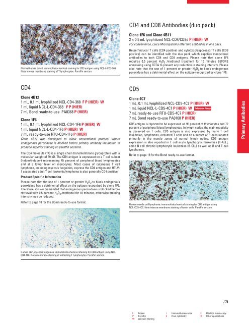
Normal human tonsil: immunohistochemical staining for CD3 antigen using NCL-L-CD3-565.<br />
Note intense membrane staining of T lymphocytes. Paraffin section.<br />
CD4<br />
Clone 4B12<br />
1 mL, 0.1 mL lyophilized NCL-CD4-368 F P (HIER) W<br />
1 mL liquid NCL-L-CD4-368 F P (HIER)<br />
7 mL Bond ready-to-use PA0368 P (HIER)<br />
Clone 1F6<br />
1 mL, 0.1 mL lyophilized NCL-CD4-1F6 P (HIER) W<br />
1 mL liquid NCL-L-CD4-1F6 P (HIER) W<br />
7 mL ready-to-use RTU-CD4-1F6 P (HIER)<br />
Clone 4B12 was developed to allow conventional protocol where<br />
endogenous peroxidase is blocked before primary antibody incubation to<br />
produce superior staining on paraffin sections.<br />
The CD4 molecule (T4) is a single chain transmembrane glycoprotein with a<br />
molecular weight of 59 kD. The CD4 antigen is expressed on a T cell subset<br />
(helper/inducer) representing 45 percent of peripheral blood lymphocytes<br />
and at a lower level on monocytes. Most cases of cutaneous T cell<br />
lymphoma, including mycosis fungoides, express the CD4 antigen and HTLV-<br />
1 associated adult T cell leukemia/lymphoma is also generally CD4 positive.<br />
Product Specific Information<br />
Please note that the use of 1 percent or greater H2O2 to block endogenous<br />
peroxidase has a detrimental effect on the epitope recognized by clone 1F6.<br />
Therefore, it is recommended that endogenous peroxidase is blocked before<br />
retrieval with 0.5 percent H2O2 /methanol for 10 minutes, otherwise staining<br />
intensity may be reduced.<br />
Refer to page 18 for the Bond ready-to-use format.<br />
Human skin, mycosis fungoides: immunohistochemical staining for CD4 antigen using NCL-<br />
CD4-1F6. Note membrane staining of infiltrating T lymphocytes. Paraffin section.<br />
CD4 and CD8 Antibodies (duo pack)<br />
Clone 1F6 and Clone 4B11<br />
2 × 0.5 mL lyophilized NCL-CD4/CD8d P (HIER) W<br />
For convenience, Leica Microsystems offer two antibodies in one pack.<br />
Helper/inducer T cells (CD4 positive) and cytotoxic/suppressor T cells (CD8<br />
positive) can be identified with the duo pack which supplies monoclonal<br />
antibodies to both CD4 and CD8 antigens. Please note that clone 1F6<br />
requires 0.5 percent H2O2 /methanol treatment for 10 minutes BEFORE<br />
unmasking using EDTA to prevent any reduction in staining intensity. Please<br />
also note that the use of 1 percent or greater H2O2 to block endogenous<br />
peroxidase has a detrimental effect on the epitope recognized by clone 1F6.<br />
CD5<br />
Clone 4C7<br />
1 mL, 0.1 mL lyophilized NCL-CD5-4C7 P (HIER) W<br />
1 mL liquid NCL-L-CD5-4C7 P (HIER) W<br />
7 mL ready-to-use RTU-CD5-4C7 P (HIER)<br />
7 mL Bond ready-to-use PA0168 P (HIER)<br />
Reference Range<br />
CD5 antigen is reported to be expressed on 95 percent of thymocytes and 72<br />
percent of peripheral blood lymphocytes. In lymph nodes, the main reactivity<br />
is observed on T cells. CD5 antigen is also expressed by many T cell<br />
leukemias, lymphomas, activated T cells and on a subset of B cells located<br />
primarily in the mantle zones of normal lymph nodes. CD5 antigen<br />
expression is also reported in T cell acute lymphocytic leukemias (T-ALL),<br />
some B cell chronic lymphocytic leukemias (B-CLL) as well as B and T cell<br />
lymphomas.<br />
Refer to page 18 for the Bond ready-to-use format.<br />
Human mantle cell lymphoma: immunohistochemical staining for CD5 antigen using<br />
NCL-CD5-4C7. Note intense membrane staining of tumor cells. Paraffin section.<br />
F Frozen I Immunofluorescence E Electron microscopy<br />
P Paraffin C Flow cytometry O Other applications<br />
W Western blotting<br />
/71<br />
Primary Antibodies


