

Labelling Review row-Online
Labelling Review row-Online
Labelling Review row-Online

Create successful ePaper yourself
Turn your PDF publications into a flip-book with our unique Google optimized e-Paper software.
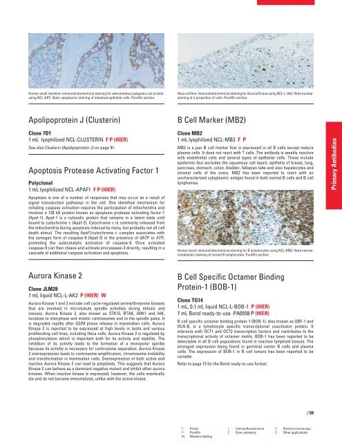
Human small intestine: immunohistochemical staining for adenomatous polyposis coli protein<br />
using NCL-APC. Note cytoplasmic staining of intestinal epithelial cells. Paraffin section.<br />
Apolipoprotein J (Clusterin)<br />
Clone 7D1<br />
1 mL lyophilized NCL-CLUSTERIN F P (HIER)<br />
See also Clusterin (Apolipoprotein J) on page 91.<br />
Apoptosis Protease Activating Factor 1<br />
Polyclonal<br />
1 mL lyophilized NCL-APAF1 F P (HIER)<br />
Apoptosis is one of a number of responses that may occur as a result of<br />
signal transduction pathways in the cell. One identified mechanism for<br />
initiating caspase activation requires the participation of mitochondria and<br />
involves a 130 kD protein known as apoptosis protease activating factor-1<br />
(Apaf-1). Apaf-1 is a cytosolic protein that remains in a latent state until<br />
bound to cytochrome c (Apaf-2). Cytochrome c is commonly released from<br />
the mitochondria during apoptosis induced by many, but probably not all cell<br />
death stimuli. The resulting Apaf1/cytochrome c complex associates with<br />
the zymogen form of caspase-9 (Apaf-3) in the presence of dATP or ATP,<br />
promoting the autocatalytic activation of caspase-9. Once activated<br />
caspase-9 can then cleave and activate procaspase-3 directly, resulting in a<br />
cascade of additional caspase activation and apoptosis.<br />
Aurora Kinase 2<br />
Clone JLM28<br />
1 mL liquid NCL-L-AK2 P (HIER) W<br />
Aurora Kinase 1 and 2 encode cell cycle-regulated serine/threonine kinases<br />
that are involved in microtubule spindle activities during mitosis and<br />
meiosis. Aurora Kinase 2, also known as STK15, BTAK, ARK1 and AIK,<br />
localizes to interphase and mitotic centrosomes and to the spindle poles. It<br />
is degraded rapidly after G2/M phase release in mammalian cells. Aurora<br />
Kinase 2 is reported to be expressed at high levels in testis and various<br />
proliferating cell lines, including HeLa cells. Aurora Kinase 2 is regulated by<br />
phosphorylation which is important both for its activity and stability. The<br />
inhibition of its activity leads to the formation of a monopolar spindle<br />
because its activity is necessary for centrosome separation. Aurora Kinase<br />
2 overexpression leads to centrosome amplification, chromosome instability<br />
and transformation in mammalian cells. Overexpression of both active and<br />
inactive Aurora Kinase 2 can lead to polyploidy. This suggests that Aurora<br />
Kinase 2 can behave as a dominant negative mutant and inhibit other aurora<br />
kinases. When inactive kinase is expressed, however, the cells eventually<br />
die and do not become immortalized, unlike with the active kinase.<br />
HeLa cell line: immunohistochemical staining for Aurora Kinase using NCL-L-AK2. Note nuclear<br />
staining of a proportion of cells. Paraffin section.<br />
B Cell Marker (MB2)<br />
Clone MB2<br />
1 mL lyophilized NCL-MB2 FP<br />
MB2 is a pan B cell marker that is expressed in all B cells except mature<br />
plasma cells. It does not react with T cells. The antibody is weakly reactive<br />
with endothelial cells and several types of epithelial cells. These include<br />
epidermis (but excludes the squamous cell layer), epithelia of breast, lung,<br />
pancreas, stomach, colon, bladder, fallopian tube and also hepatocytes and<br />
stromal cells of the ovary. MB2 has been reported to react with an<br />
uncharacterized cytoplasmic antigen found in both normal B cells and B cell<br />
lymphomas.<br />
Human tonsil: immunohistochemical staining for B lymphocytes using NCL-MB2. Note intense<br />
cytoplasmic staining of normal B lymphocytes. Paraffin section.<br />
B Cell Specific Octamer Binding<br />
Protein-1 (BOB-1)<br />
Clone TG14<br />
1 mL, 0.1 mL liquid NCL-L-BOB-1 P (HIER)<br />
7 mL Bond ready-to-use PA0558 P (HIER)<br />
B cell specific octamer binding protein-1 (BOB-1), also known as OBF-1 and<br />
OCA-B, is a lymphocyte specific transcriptional coactivator protein. It<br />
interacts with OCT1 and OCT2 transcription factors and contributes to the<br />
transcriptional activity of octamer motifs. BOB-1 has been reported to be<br />
detectable in all B cell populations found in reactive lymphoid tissues. The<br />
strongest expression being found in germinal center B cells and plasma<br />
cells. The expression of BOB-1 in B cell tumors has been reported to be<br />
variable.<br />
Refer to page 15 for the Bond ready-to-use format.<br />
F Frozen I Immunofluorescence E Electron microscopy<br />
P Paraffin C Flow cytometry O Other applications<br />
W Western blotting<br />
/59<br />
Primary Antibodies


