

Labelling Review row-Online
Labelling Review row-Online
Labelling Review row-Online

Create successful ePaper yourself
Turn your PDF publications into a flip-book with our unique Google optimized e-Paper software.
Primary Antibodies<br />
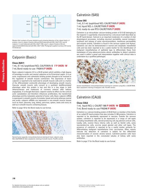
Western blot: analysis of human skeletal muscle showing detection of the calpain family of<br />
proteins. Lane A, calpain 3 bands at 94 and 30 kD detected with NCL-CALP-2C4. Lane B,<br />
calpains 1, 2 and 3 detected with NCL-CALP-11B3. Lane C, calpain 3 bands at 94 and<br />
approximately 60 kD detected with NCL-CALP-12A2. Photograph supplied courtesy of Dr Louise<br />
V B Anderson.<br />
Calponin (Basic)<br />
Clone 26A11<br />
1 mL, 0.1 mL lyophilized NCL-CALPONIN-B F P (HIER) W<br />
7 mL Bond ready-to-use PA0416 P (HIER)<br />
Basic calponin (calponin-h1) is a 34 kD protein which exhibits a high degree<br />
of homology to acidic and neutral calponins at its N-terminal region. It is an<br />
actin, tropomyosin and calmodulin binding protein thought to be involved in<br />
the regulation of smooth muscle contraction. The expression of basic<br />
calponin is reported to be restricted to smooth muscle cells and is a marker<br />
of the differentiated contractile phenotype of developing smooth muscle.<br />
Vascular smooth muscle cells convert to a synthetic dedifferentiated<br />
phenotype when this protein is lost and this is a key stage in both<br />
atherosclerosis and restenosis of coronary arteries after balloon<br />
angioplasty. It is thought that basic calponin exerts its effect via the cortical<br />
actin cytoskeleton and therefore influences proliferation, the transformed<br />
phenotype and the metastatic potential of tumor cells. Basic calponin mRNA<br />
is expressed in smooth muscle of prostate, bowel and aorta whereas neutral<br />
and acidic calponin mRNAs are expressed in non-smooth muscle tissues<br />
such as heart, placenta, lung, kidney, pancreas, spleen, testis and ovary as<br />
well as in smooth muscle-containing tissues.<br />
Refer to page 16 for the Bond ready-to-use format.<br />
Normal human appendix: immunohistochemical staining for basic calponin using<br />
NCL-CALPONIN-B. Note cytoplasmic staining of smooth muscle. Paraffin section.<br />
/66<br />
For detailed information on all products please visit our website:<br />
www.leica-microsystems.com<br />
Calretinin (5A5)<br />
Clone 5A5<br />
1 mL, 0.1 mL lyophilized NCL-CALRETININ P (HIER)<br />
1 mL liquid NCL-L-CALRETININ P (HIER)<br />
7 mL ready-to-use RTU-CALRETININ P (HIER)<br />
Calretinin is an intracellular calcium-binding protein of 31.5 kD belonging to<br />
the troponin C superfamily characterized by a structural motif described as<br />
the EF-hand domain. Calcium is an important moderator of a number of vital<br />
physiological processes, including neuronal excitability, axonal transport,<br />
synthesis and release of some neurotransmitters, membrane permeability<br />
and enzyme activity. Calretinin is found in the nervous system and thymus.<br />
Calretinin can also be demonstrated in normal and neoplastic mesothelial<br />
cells and has been reported to be a useful marker for the identification of<br />
malignant mesotheliomas of epithelial type to differentiate these from<br />
metastases of lung adenocarcinoma where antibodies to detect calretinin<br />
are used in within a panel and interpretation together with clinical data is<br />
undertaken by a qualified pathologist.<br />
Human mesothelioma: immunohistochemical staining for calretinin using NCL-L-CALRETININ.<br />
Note cytoplasmic staining of malignant cells. Paraffin section.<br />
Calretinin (CAL6)<br />
Clone CAL6<br />
1 mL liquid NCL-L-CALRET-566 P (HIER) W<br />
7 mL Bond ready-to-use PA0346 P (HIER)<br />
Calretinin is a calcium-binding protein of 29 kD that is a member of the family<br />
of so-called EF-hand proteins that also includes S-100 proteins. Calretinin is<br />
reported to be abundantly expressed in neurons. Outside the nervous<br />
system, calretinin is reported to be expressed in a range of cell types<br />
including mesothelial cells, steroid producing cell, (eg adrenal cortical cells,<br />
Leydig cells, ovarian theca interna cells as well as Sertoli cells, some<br />
neuroendocrine cells, eccrine sweat glands) and other cell types. The<br />
presence of calretinin is reported to be a useful marker primarily for<br />
differentiating malignant mesothelioma from carcinomas. Other reports<br />
indicate the detection of calretinin is helpful for the differential<br />
characterization of ovarian stroma tumors. Calretinin-positive cells have<br />
also been reported in the convoluted tubules of kidney with some<br />
antibodies.<br />
Refer to page 17 for the Bond ready-to-use format.<br />
Products in this catalog are subject to regulatory approval.<br />
This catalog is not for use in the USA.<br />
Reference Range


