

Labelling Review row-Online
Labelling Review row-Online
Labelling Review row-Online

You also want an ePaper? Increase the reach of your titles
YUMPU automatically turns print PDFs into web optimized ePapers that Google loves.
Primary Antibodies<br />
Motility-Related Protein-1 (CD9)<br />
Clone 72F6<br />
1 mL lyophilized NCL-CD9 F P (HIER)<br />
See also CD9 (Motility-Related Protein-1) on page 72.<br />
Muc Glycoprotein Antibodies<br />
Clone Ma552<br />
1 mL lyophilized muc-1 core glycoprotein<br />
NCL-MUC-1-CORE F P (HIER)<br />
Clone Ma695<br />
1 mL lyophilized muc-1 glycoprotein<br />
NCL-MUC-1 F P (HIER)<br />
Clone Ccp58<br />
1 mL, 0.1 mL lyophilized muc-2 glycoprotein<br />
NCL-MUC-2 F P (HIER)<br />
Clone CLH2<br />
1 mL, 0.1 mL lyophilized muc-5AC glycoprotein<br />
NCL-MUC-5AC P (HIER)<br />
Clone CLH5<br />
1 mL, 0.1 mL lyophilized muc-6 glycoprotein<br />
NCL-MUC-6 P (HIER)<br />
Mucins are heavily glycosylated proteins which constitute the major<br />
components of mucus covering the surface of epithelial tissues. Nine<br />
distinct epithelial mucin genes (Muc-1, 2, 3, 4, 5AC, 5B, 6, 7 and 8) have been<br />
identified. Various immunohistochemical and in situ hybridization studies<br />
have reported that these mucins are differentially expressed in epithelia<br />
with cell-type specificity. The normal gastric mucosa shows cell-type<br />
specific expression of Muc-1, Muc-5AC and Muc-6 glycoproteins. Muc-1<br />
and Muc-5AC are found in superficial epithelium and Muc-6 glycoprotein in<br />
the deep glands. Muc-1 and Muc-5AC glycoproteins are reported to be<br />
expressed in many epithelia but Muc-6 glycoprotein is mainly expressed in<br />
gastric mucosa. In addition, Muc-2 glycoprotein is not expressed in normal<br />
gastric mucosa. In gastric cancer, alterations in mucin polypeptide<br />
expression have been reported, including the loss of expression of Muc-5AC<br />
glycoprotein, increased mucin heterogeneity, glycosylation changes and the<br />
expression of simple mucin-type carbohydrates.<br />
Normal human stomach: immunohistochemical staining for Muc-6 glycoprotein using NCL-<br />
MUC-6. Note cytoplasmic staining of mucus secreting cells of the deep glands. Paraffin<br />
section.<br />
/ 136<br />
For detailed information on all products please visit our website:<br />
www.leica-microsystems.com<br />
Multiple Myeloma Oncogene 1 (MUM-1)<br />
Clone EAU32 New!<br />
1 mL, 0.1 mL liquid NCL-L-MUM1 P (HIER)<br />
7 mL Bond ready-to-use PA0129 P (HIER)<br />
The MUM-1 (multiple myeloma oncogene 1) gene was originally identified<br />
because of it’s involvement in the t(6:14) translocation observed in multiple<br />
myeloma, which causes the juxtaposition of the MUM-1 gene to the Ig heavy<br />
chain locus. MUM-1 is expressed in late plasma cell directed stages of<br />
B cell differentiation and in activated T cells, suggesting that MUM-1 may<br />
serve as a marker for lympho-hemopoetic neoplasms derived from these<br />
cells. The morphologic spectrum of MUM-1 expressing cells has been found<br />
to range from that of a centrocyte to that of a plasmablast/plasma cell.<br />
Consequently the histogenic value of MUM-1 may be to provide a marker to<br />
aid in the identification of the transition from BCL-6 positive (germinal center<br />
B cells) to CD138 positive (immunoblasts and plasma cells). MUM-1<br />
expression occurs in a wide range of lymphoid neoplasms including a<br />
proportion of diffuse B cell lymphomas but not myeloid or extra-hemopoietic<br />
neoplasms. MUM-1 is consistently expressed in myeloma cells, Reed<br />
Sternberg cells in classic Hodgkin Disease, and activated and neoplastic<br />
T cells.<br />
Refer to page 35 for the Bond ready-to-use format.<br />
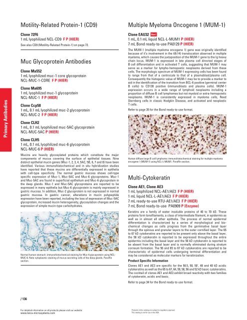
Human diffuse large B cell lymphoma: immunohistochemical staining for multiple myeloma<br />
oncogene 1 (MUM-1) using NCL-L-MUM1. Paraffin section.<br />
Multi-Cytokeratin<br />
Clone AE1, Clone AE3<br />
1 mL lyophilized NCL-AE1/AE3 F P (HIER)<br />
1 mL liquid NCL-L-AE1/AE3 F P (HIER)<br />
7 mL ready-to-use RTU-AE1/AE3 F P (HIER)<br />
7 mL Bond ready-to-use PA0909 P (Enzyme)<br />
Keratins are a family of water insoluble proteins of 40 to 70 kD. These<br />
proteins form tonofilaments, a class of intermediate filament, in epidermis as<br />
well as in almost all other epithelia. The process of normal epidermal<br />
differentiation is characterized by a series of morphological and biochemical<br />
changes as cells progress from the germinative basal layer<br />
through the spinous and granular layers to the outer cornified layer. The 65<br />
to 67 kD cytokeratins are reported to be present only above the basal layer,<br />
the 58 kD cytokeratin is reported to be expressed throughout the entire<br />
epidermis including the basal layer and the 56 kD cytokeratin is reported to<br />
be absent from the basal layer and is normally eliminated during stratum<br />
corneum formation. The 56 and 65 to 67 kD cytokeratins are reported to be<br />
characteristic of epidermal cells undergoing terminal differentiation and<br />
may be considered as molecular markers for keratinization.<br />
Product Specific Information<br />
Clones AE1 and AE3 are specific for the 56.5, 50, 50', 48 and 40 kD acidic<br />
cytokeratins as well as the 65 to 67, 64, 59, 58, 56 and 52 kD basic cytokeratins.<br />
The cocktail of clones AE1 and AE3 exhibit broad reactivity with two families<br />
of cytokeratin, acidic and basic.<br />
Refer to page 34 for the Bond ready-to-use format.<br />
Products in this catalog are subject to regulatory approval.<br />
This catalog is not for use in the USA.


